Download
1 / 14
140 likes | 212 Views
Justin Williams, PhD Department of Biomedical Engineering University of Wisconsin-Madison. Fredrick Kelcz, PhD, MD Department of Radiology University of Wisconsin-Madison. Advisor Client. Abstract.

E N D
Justin Williams, PhD Department of Biomedical Engineering University of Wisconsin-Madison Fredrick Kelcz, PhD, MD Department of Radiology University of Wisconsin-Madison Advisor Client
Abstract Core biopsy procedures are currently subject to high specificity due to inaccurate imaging of small tumors around the biopsy needle. Ultrasonic imaging can differentiate between tissue types by the differences in echogenic calcifications. The basics of IVUS probes were used to develop two ultrasonic methods: an integrated ultrasonic probe and a catheter based IVUS probe technique. The chosen design involves the insertion of a catheter based IVUS probe, once the needle is in place. This technique should provide accurate imaging and is extremely practical to implement. However, this techniques will require further experimentation to evaluate its ability to image within the needle in the presence of a strong magnetic field.
Problem Statement The goal of this project is to develop an intravascular ultrasonic probe (IVUS) to provide better imaging without artifact during mammographic and MRI guided biopsy procedures. Usually Biopsy procedures are done with MRI or CT guidance; however, biopsy needles can obstruct imaging of tumors making it difficult to make fine adjustments in needle placement.
Problem Definition • Motivation The accuracy of the overall biopsy procedure is improved and doctors can guarantee patients that the correct lesion was sampled
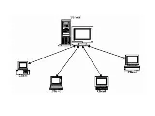
Background on Guided Core Biopsy Procedure • First visit • Initial mammographic/MR image • Positive diagnosis—suspicious lesions • Second visit • Second MR image—insertion of plastic clip • Third MR image—check placement • Needle Insertion • Exact location never verified • Perform biopsy • Results • Pathological analysis
IVUS Probes • Use a pizoelectric transducer implanted in catheter 1-2mm in diameter and 100-130cm long • Provide 2D cross sectional imaging • A motor rotates the transducer at ~1800rpm • Typically single use costing ~$1000 • Operate at 20-40MHz depending on model • Imaging depth of 1-2cm depending on frequency
Design Criteria • Spatial resolution approximately 0.5-1mm • Depth perception approximately 3-5mm • Distinguish small tumors and calcifications from normal tissue • Operate within core biopsy needle with minimal artifact • MRI compatible (Class III)
Final Design: Catheter Based IVUS Probe • 40 MHz “Discovery” IVUS probe • Core Biopsy Needle • Manufactured plastic rod insert
Testing Methods • Phantoms • Water • Ground orange pulp* • Orange slice* • Ultrasonic gel* • Raw turkey breast* *With and without simulated calcifications. To simulate calcifications we used 8 in 1:Sun ripened fruit mineral treat (22.5-27% calcium content)
Future Work • Testing MRI compatibility • Future phantoms • Simulated tumor (olive in turkey breast) • Real specimen • Investigate different probe frequencies • Investigate probe redesign
Conclusions • IVUS imaging shows great potential towards increasing biopsy yields • System is capable of detecting calcifications • Ability to detect actual tumors is unknown • Current devices are not MRI compatible
Acknowledgements Dr. Frederick Kelcz Professor Justin Williams Dr. Tomy Varghese Dr. Jim Zagzebski Keith Bourne Our team would like to extend thanks to the following individuals for their assistance on this design project:
References Diagnosis by Biopsy. 2003. The Breast Center at Southern Illinois University School of Medicine. Date Accessed: February 9, 2004. URL: http://www.siumed.edu/breastcenter/diagnosis.html. Lengyel J, Greenberg DP, and Popp R. Time-Dependent Three-Dimensional intravascular ultrasound. SIGGRAPH 95 Conference Proceedings, Annual Conference Series, pages 457--464. ACM SIGGRAPH, Addison Wesley, August 1995 Nissen S. 2001.Coronary angiography and intravascular ultrasound. Am J Cardiol. 87(4A):15A-20A Trevino, M. (2003, December 12) Calcifications may be presage invasive breast cancer. Diagnostic Imaging Online. Retrieved February 11, 2004 from http://www.diagnosticimaging.com/dinews/2003121201.shtml Yock, P., Johnson, E., and David, D. Intravascular ultrasound:Development and clinical potential. American Journal of CardiacImaging 2(185)