Download
1 / 1
110 likes | 597 Views
The MRI Study of Normal Brain Development. AMYGDALA VOLUME IS ASSOCIATED WITH SUBCLINICAL ANXIOUS/DEPRESSED PROBLEMS AMONG TYPICALLY DEVELOPING YOUTHS. Albaugh , M.D., 1 Karama , S., 2 Collins, D.L., 2 Ducharme , S., 2 Botteron , K.N., 3 Evans, A.C., 2

E N D
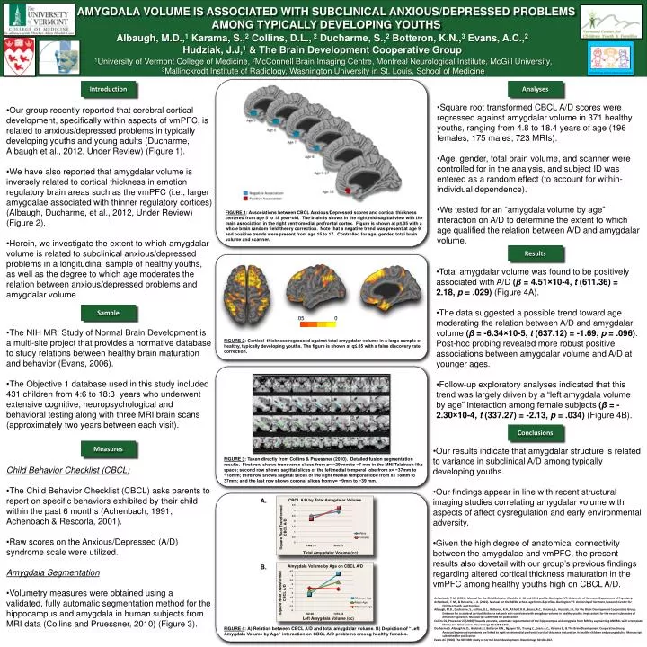
The MRI Study of Normal Brain Development AMYGDALA VOLUME IS ASSOCIATED WITH SUBCLINICAL ANXIOUS/DEPRESSED PROBLEMS AMONG TYPICALLY DEVELOPING YOUTHS Albaugh, M.D.,1Karama, S.,2 Collins, D.L., 2Ducharme, S.,2Botteron, K.N.,3 Evans, A.C.,2 Hudziak, J.J,1 & The Brain Development Cooperative Group 1University of Vermont College of Medicine, 2McConnell Brain Imaging Centre, Montreal Neurological Institute, McGill University, 3Mallinckrodt Institute of Radiology, Washington University in St. Louis, School of Medicine Introduction Analyses • Our group recently reported that cerebral cortical development, specifically within aspects of vmPFC, is related to anxious/depressed problems in typically developing youths and young adults (Ducharme, Albaugh et al., 2012, Under Review) (Figure 1). • We have also reported that amygdalar volume is inversely related to cortical thickness in emotion regulatory brain areas such as the vmPFC (i.e., larger amygdalae associated with thinner regulatory cortices) (Albaugh, Ducharme, et al., 2012, Under Review) (Figure 2). • Herein, we investigate the extent to which amygdalar volume is related to subclinical anxious/depressed problems in a longitudinal sample of healthy youths, as well as the degree to which age moderates the relation between anxious/depressed problems and amygdalar volume. • Square root transformed CBCL A/D scores were regressed against amygdalar volume in 371 healthy youths, ranging from 4.8 to 18.4 years of age (196 females, 175 males; 723 MRIs). • Age, gender, total brain volume, and scanner were controlled for in the analysis, and subject ID was entered as a random effect (to account for within-individual dependence). • We tested for an “amygdala volume by age” interaction on A/D to determine the extent to which age qualified the relation between A/D and amygdalar volume. FIGURE 1: Associations between CBCL Anxious/Depressed scores and cortical thickness centered from age 5 to 18 year-old. The brain is shown in the right mid-sagittal view with the main association in the right ventromedial prefrontal cortex. Figure is shown at p≤.05 with a whole brain random field theory correction. Note that a negative trend was present at age 9, and positive trends were present from age 15 to 17. Controlled for age, gender, total brain volume and scanner. Results • Total amygdalar volume was found to be positively associated with A/D (β = 4.51×10⁻⁴, t (611.36) = 2.18, p = .029) (Figure 4A). • The data suggested a possible trend toward age moderating the relation between A/D and amygdalar volume (β = -6.34×10⁻⁵, t (637.12) = -1.69, p = .096). Post-hoc probing revealed more robust positive associations between amygdalar volume and A/D at younger ages. • Follow-up exploratory analyses indicated that this trend was largely driven by a “left amygdala volume by age” interaction among female subjects (β = -2.30×10⁻⁴, t (337.27) = -2.13, p = .034) (Figure 4B). Sample • The NIH MRI Study of Normal Brain Development is a multi-site project that provides a normative database to study relations between healthy brain maturation and behavior (Evans, 2006). • The Objective 1 database used in this study included 431children from 4:6 to 18:3 years who underwent extensive cognitive, neuropsychological and behavioral testing along with three MRI brain scans (approximately two years between each visit). FIGURE 2: Cortical thickness regressed against total amygdalar volume in a large sample of healthy, typically developing youths. The figure is shown at q≤.05 with a false discovery rate correction. Conclusions Measures • Our results indicate that amygdalar structure is related to variance in subclinical A/D among typically developing youths. • Our findings appear in line with recent structural imaging studies correlating amygdalar volume with aspects of affect dysregulation and early environmental adversity. • Given the high degree of anatomical connectivity between the amygdalae and vmPFC, the present results also dovetail with our group’s previous findings regarding altered cortical thickness maturation in the vmPFC among healthy youths high on CBCL A/D. • Child Behavior Checklist (CBCL) • The Child Behavior Checklist (CBCL) asks parents to report on specific behaviors exhibited by their child within the past 6 months (Achenbach, 1991; Achenbach & Rescorla, 2001). • Raw scores on the Anxious/Depressed (A/D) syndrome scale were utilized. • Amygdala Segmentation • Volumetry measures were obtained using a validated, fully automatic segmentation method for the hippocampus and amygdala in human subjects from MRI data (Collins and Pruessner, 2010) (Figure 3). FIGURE 3: Taken directly from Collins & Pruessner (2010). Detailed fusion segmentation results. First row shows transverse slices from z= −29 mm to −7 mm in the MNI Talairach-like space; second row shows sagittal slices of the leftmedial temporal lobe from x= −37mm to −18mm; third row shows sagittal slices of the right medial temporal lobe from x= 18mm to 37mm; and the last row shows coronal slices from y= −9mm to −39 mm. A. B. .05 0 Achenbach, T. M. (1991). Manual for the Child Behavior Checklist 4–18 and 1991 profile. Burlington VT: University of Vermont, Department of Psychiatry. Achenbach, T. M., & Rescorla, L. A. (2001). Manual for the ASEBA school-age forms & profiles. Burlington VT: University of Vermont, Research Center for Children,Youth, and Families. Albaugh, M.D., Ducharme, S., Collins, D.L., Botteron, K.N., Althoff, R.R., Evans, A.C., Karama, S., Hudziak, J.J., for the Brain Development Cooperative Group. Evidence for a cerebral cortical thickness network anti-correlated with amygdalar volume in healthy youths: implications for the neural substrates of emotion regulation. Manuscript submitted for publication. Collins DL, Pruessner JC (2010) Towards accurate, automatic segmentation of the hippocampus and amygdala from MRI by augmenting ANIMAL with a template library and label fusion. Neuroimage 52:1355-1366. Ducharme S. Albaugh M.D., Hudziak J.J, Botteron K.N., Nguyen T.V., Truong C., Evans A.C., Karama S., & The Brain Development Cooperative Group. Anxious/depressed symptoms are linked to right ventromedial prefrontal cortical thickness maturation in healthy children and young adults. Manuscript submitted for publication. Evans AC (2006) The NIH MRI study of normal brain development. Neuroimage 30:184-202. FIGURE 4: A) Relation between CBCL A/D and total amygdalar volume. B) Depiction of “Left Amygdala Volume by Age” interaction on CBCL A/D problems among healthy females.