Download
1 / 57
650 likes | 1.02k Views
Medical Retina and Macular Diseases. Dr. Timothy Y. Y. Lai MBBS, MMedSc, MRCSEd, FCOphthHK, FHKAM(Ophth) Department of Ophthalmology and Visual Sciences The Chinese University of Hong Kong. Medical Retina.

E N D
Medical Retina and Macular Diseases Dr. Timothy Y. Y. Lai MBBS, MMedSc, MRCSEd, FCOphthHK, FHKAM(Ophth) Department of Ophthalmology and Visual Sciences The Chinese University of Hong Kong
Medical Retina • A specialty that deals with the investigation and non-surgical treatment of retinal disorders • Retinal diseases associated with systemic diseases • Diabetic Retinopathy • Hypertensive Retinopathy • Vascular retinopathies • Medical macular diseases • Age-related Macular Degeneration
Diabetic Retinopathy • One of the leading causes of blindness • Risk factors • Duration of diabetes • 80% of type I and 70% of type II diabetics have retinopathy after 15 yrs • Type of diabetes mellitus • Control of hyperglycemia • Hypertension • Associated renal disease • Pregnancy
Diabetic Retinopathy • Classification • Non-proliferative (NPDR) • Mild • Moderate • Severe • Proliferative (PDR)
Non-proliferative Diabetic Retinopathy (NPDR) • Pathogenesis • Microvascular disease causing capillary damage • Leakage of blood constituents into the retina • Retinal hemorrhages • Retinal edema • Lipid exudation
Non-proliferative Diabetic Retinopathy (NPDR) • Dot and blot hemorrhage • Hard exudate • Cotton-wool spots • Venous beading • Venous loops
Proliferative Diabetic Retinopathy (PDR) • Pathogenesis • Retinal ischemia causing neovascularization • May be asymptomatic if only neovascularization without hemorrhage
Neovascularization at Disc (NVD) Neovascularization elsewhere (NVE)
Causes of Visual Loss in DR • Macular Edema • Complications of PDR • Vitreous hemorrhage • Fibrous tissue proliferation • Retinal detachment
Microaneurysms Circinate exudate Retinal edema
Vitreous Hemorrhage Tractional Retinal Detachment Combined Retinal Detachment
Diabetic Retinopathy • Treatment • Laser photocoagulation • Focal or grid: for macular edema • Pan-retinal photocoagulation: for PDR • Control of systemic disease • Hyperglycemia • Hypertension • Renal disease • Vitreous surgery
Laser Photocoagulation Outpatient procedure Topical Anesthesia Multiple Sessions In PDR, laser should be performed before vitreous hemorrhage and retinal detachment develops
Diabetic Retinopathy • Early identification of the disease and prompt referral to the ophthalmologist • Dilate your patients for examination with ophthalmoscope regularly • Prompt treatment reduces risk of visual loss by 50% • Patients may be asymptomatic but still have advanced PDR
Hypertensive Retinopathy • Focal or generalized narrowing of retinal arteries associated with hypertension • Clinical features • Cotton-wool spots • Hard exudates • Macular star • Macular edema • Retinal hemorrhage • Optic disc swelling
Severe Hypertensive Retinopathy Macular star Disc swelling
Hypertensive Retinopathy • Management • Rule out secondary hypertension • Control of hypertension
Retinal Vascular Occlusions • Venous occlusion more common than arterial occlusion • Pathogenesis • Arterial occlusion – embolus • Central retinal artery occlusion (CRAO) • Branch retinal artery occlusion (BRAO) • Venous occlusion – abnormal blood flow • Central Retinal Vein Occlusion (CRVO) • Branch Retinal Vein Occlusion (BRVO)
Retinal Arterial Occlusions • Symptoms • Sudden, painless, marked loss of vision • Immediate treatment within 24-48 hours may be beneficial in some patients • Systemic Associations • Cardiovascular disease • Carotid artery disease • Temporal arteritis / inflammatory arteritis • Coagulopathies
Central Retinal Artery Occlusion Afferent Pupillary Defect Cherry Red Spot Retinal Edema Branch Retinal Artery Occlusion
Retinal Venous Occlusions • Symptoms • Sudden painless loss of vision • Various extent of visual loss • Systemic Associations • Diabetes Mellitus • Hypertension • Hematological diseases • Vasculitis
Central Retinal Vein Occlusion Branch Retinal Vein Occlusion
Neovascular glaucoma Macular edema Laser photocoagulation
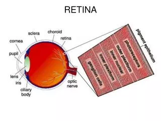
Where is the macula? 脈絡膜 Choroid 鞏膜 Sclera 視網膜 Retina 角膜 Cornea 黃斑區 Macula 晶體 Lens Iris 虹膜 視神經 Optic nerve Cilliary body 睫狀體
Fovea Macula
Macular Diseases • Common surgical macular diseases • Macular hole • Epiretinal membrane • Common medical macular diseases • Age-related macular degeneration (AMD) • Myopic maculopathy • Central serous chorioretinopathy (CSC)
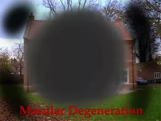
Age-related Macular Degeneration (AMD) • Leading cause of severe vision loss in people > 50 years in the western world • Visual loss due to drusens / RPE degeneration or development choroidal neovascularization (CNV)
Age-related Macular Degeneration (AMD) • Two forms • Dry (non-neovascular) AMD(80% to 90%) • Wet (neovascular) AMD(10% to 20%) • 90% of vision loss is caused by wet form of AMD
Symptoms of AMD – Early Decrease in color and contrast sensitivity
Symptoms of AMD – Intermediate Impairment of central visual function
Symptoms of AMD – Intermediate Metamorphopsia, distortion of central image
Symptoms of AMD – Late Central Scotoma
Hallmark of AMDDevelopment of Drusen Bruch’sMembrane Drusen
Early Dry AMD • Asymptomatic • Examination reveals several small drusen or a few medium-sized drusen (63-124m)
Intermediate Dry AMD • Many medium-sized drusen or 1 large drusen (>125m) • Vision may be impaired
Advanced Dry AMD • More severe visual impairment • Presence of drusen with degeneration of RPE • Geographic atrophy
Dry AMD Wet AMDFormation of New Vessels ChoroidalNeovascularization
Diagnosis • Vision function testing • Visual acuity • Amsler grid • Ophthalmolscopy • Fluorescein angiography
Early Diagnosis • Amsler Grid • Adequate lighting • Wear reading glasses • Hold the Amsler grid at normal reading distance (about 30cm) • Cover one eye at a time • Stare at the center dot • Ask the following questions: • Are any of the lines wavy, missing, blurry, or discolored? • Are any of the boxes different in size or shape from the others? 5mm squares 10 cm x 10 cm
Self monitoring with Amsler Grid Normal Abnormal
Fluorescein angiography Intravenous injection Uptake of fluorescein dye at the site of abnormal vessels - Size, Location, Activity
Management of AMD • Treatment for neovascular AMD • Laser photocoagulation • Submacular surgery • Photodynamic therapy (PDT) with Verteporfin • Anti-angiogenesis therapy (Anti-VEGF) • Prevent progression to advanced AMD • Antioxidants • Quit smoking • Low-vision aids
Laser Photocoagulation • Non-selective thermal laser photocoagulation • Destroy CNV • Irreversible damage to the overlying retina and RPE • Side effects of immediate scotoma or drop in central vision • CNV persist or recur in 50% of patients