Download

1 / 36
360 likes | 499 Views
Manzano, Clairol Marcelo, Pamela Marcial, Karmi Margaret Matematico, Michelle Matias, Evangelyn Maulion, Marienelle. Benign Breast Mass. Three females with ages 23, 35, and 55 years, respectively, went to see you for consult. All have a mass in one of their breasts. .

E N D
Manzano, Clairol Marcelo, Pamela Marcial, Karmi Margaret Matematico, Michelle Matias, Evangelyn Maulion, Marienelle Benign Breast Mass
Three females with ages 23, 35, and 55 years, respectively, went to see you for consult. All have a mass in one of their breasts.
What important general data from the patients do you think are important to be able to guide you in your diagnosis? Explain. • Breast Lump • Age • Family History • Reproduction and Menstrual History • Radiation Exposure • Hormone Replacement Therapy • Oral Contraceptives • Body Mass Index
Breast Lump • Time of recognition • Number • Size • Changes before menstruation • Location • Shape • Borders • Mobility • Tenderness • Pain
Risk Factors • Age = Breast Cancer risk • Family History • Relative Risk of Cancer: 1st degree > 2nd degree Sister > Mother Highest risk: (+) FH – Mother and Sister • Early Menarche & Late Menopause • Breast Cancer risk • Nulligravidity is a risk • Radiation Exposure = Breast Cancer risk • HRT, Contraceptives, Obesity • Estrogen Exposure = Breast Cancer risk
How will you approach the 23-year old, with a 2 X 2 X 2cm, firm, mobile, well-circumscribed non-tender mass on her L breast?
Diagnosis • FIBROADENOMA • Benign fibroepithelial neoplasm from the terminal duct lobular unit of the breast • Painless, firm, solitary, mobile • Occurs in young women of child bearing years • (20 to 30 years old) • Size: 2 – 3 cm • Borders: Well-defined • Margins: Regular • NOT PREMALIGNANT
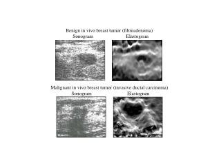
Role of Imaging Modalities • Mammography • Indeterminate Mass with suspicion of cancer • Pre-requisite prior BCT • Follow-up after BCT and of contralateral breast • Ultrasound • Distinguish between solid and cystic masses • Provide guidance for cyst aspiration or core biopsy • For palpable mass that is partially or poorly seen on a mammogram
Management • Fine Needle Aspiration Biopsy • Below 25 years old: OBSERVATION • 25 to 35 years old: OBSERVATION/EXCISION • Above 35 years old: EXCISION • Follow up: PE and Mammography • Cryoablation Treatment • Alternative to open surgical removal of fibroadenoma
How will you approach the 35-year old, with a 2 x 2 x 2cm, firm, mobile, well-circumscribed non-tender mass on her R breast?
Approach to the Patient • History • Physical Examination • Bilateral Mammography • Fine Needle Aspiration Biopsy • Cytology
A mammogram was taken as seen in the picture. Is this benign or malignant?
Should the patient have a mother who is a breast cancer survivor, how would that information change your management
How will you approach the 55-year old menopausal patient with a 2-cm diameter, mobile, firm, non-tender mass on her R breast?
FNAC reveals NEGATIVE FOR MALIGNANT CELLS. How would you now manage the patient?
A 43-year old female consults because of a rapidly growing L breast. Axilla is negative for clinically palpable nodes.
A 55-year old female consults because of bloody nipple discharge.
Differentiate a physiologic from pathologic nipple discharge.
Two ladies aged 20 and 48 years, respectively, consulted because of bilateral breast tenderness.
How do you differentiate the diagnosis in #1 from that of #2?
The 48-year old undergoes surgery showing the gross finding below, What is your treatment?