Download
1 / 39
390 likes | 590 Views
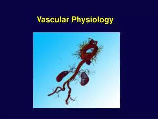
Vascular Physiology 3. Upper and lower extremity arterial conditions other than atherosclerosis. Upper extremity ischemia . Emboli. Heart most likely source of non-atherosclerotic emboli. 10-20% of all cardiac emboli lodge in upper extremity. 70% of all upper ext emboli come from heart.

E N D
Vascular Physiology 3 • Upper and lower extremity arterial conditions other than atherosclerosis.
Emboli • Heart most likely source of non-atherosclerotic emboli. 10-20% of all cardiac emboli lodge in upper extremity. 70% of all upper ext emboli come from heart. • Thrombus • Tumor • Valvular lesions • Fairly common
Raynaud’s Disease(Cold sensitivity) • Female- most frequent 18-30 years • Abnormal vasoconstriction of extremities upon exposure to cold or emotional stress. • Intermittent attacks of pallor, cyanosis,then rubor of digits (usually upper), bilateral or symetrical, normal radial and ulnar pulse. • No evidence of obstructive disease. • Fairly common
Raynaud’s Disease cont. • Treatment • Warmth, gloves, socks, avoid cold • Vasodilators
Raynaud’s Phenomenon(cold sensitivity) • Intermittent pallor, cyanosis, redness,normal. Repeats. • Response to cold or emotion. • Numbness, tingling, burning may occur. • Secondary to such conditions as occlusive arterial disease, systemic scleroderma, thoracic outlet syndrome, pulmonary hypertension, myxedema or trauma. • Fairly common
Raynaud’s Phenomenon cont. • Vascular Lab to look for underlying cause of vasoconstriction. • Vascular Lab to document vasospasm.
Arterial/Venous fistulae • Surgically constructed for hemodialysis • Cimino-Brescia: end to end or side to side anastomosis between the radial artery and cephalic vein at wrist. • Prosthetic graft (PTFE) • Loop between brachial art and antecubital vein • Straight between radial art at wrist and antecubital vein • Straight between brachial artery and subclavian vein • Common
Buerger’s Disease • Thromboangiitis Obliterans (fairly uncommon) • Men <40yrs • 99% smoke • Affects small and medium arteries, can affect veins also. • Inflammation leading to formation of thrombi • Tissue necrosis develops early because of poor collaterals in end arteries of fingers and toes.
Trauma • Dissection, thrombosing, Arterial/venous fistulae. • Acute ischemia • Can happen to upper or lower ext. • Fairly common
Thoracic Outlet Syndrome • Compression of nerve, artery, or vein in the thoracic outlet area. • Area of 1st rib, clavicle, and scalene muscle. • A “cervical rib” with or without a fibrous band may be present. • Uncommon
Types of Thoracic Outlet Syndrome. True neurogenic: Wasting of muscles of hand and hand weakness, with positive electromyography. Vascular Thoracic Outlet: an arterial or venous lesion is present on angiography. Duplex and photocell exam is helpful and most likely ordered before angio.
Thoracic Outlet Types cont. • Disputed Thoracic Outlet. • Weakness, parathesia, pain of hand, arm, shoulder girdle, chest wall, and headache. • Eletromyographic test normal. • Hand wasting is never found. • Patients do not progress to true neurogenic or vascular thoracic outlet.
Tumor • Muscular masses can be present in the upper extremity. Some are vascular and cause bruit. • Masses can compress artery or vein. • Uncommon
Rheumatic/Autoimmune Diseases Giant Cell Arteritis: Temporal arteritis and Takayasu’s Can cause arm claudication or Raynaud’s phenomenon. Uncommon
Radiation arteritis • Inflammation of subclavian and axillary arteries resulting from radiation treatment. • Uncommon but is seen
Fibromuscular dysplasia • Systemic disorder, smooth muscle hyperplasia, and general disorganization of the arterial wall. • Can cause arm claudication. Uncommon
Summary upper • Common causes for upper art conditions • Emboli • Cold sensitivity (vasospasm in Raynaud’s) • A-v grafts
Upper summary cont. • Somewhat common • Buerger’s Disease (men more than women) • Trauma
Upper summary cont. • Uncommon upper ischemia causes • Thoracic outlet • Tumor • Rheumatic/Autoimmune disease • Radiation arteritis • Fibromuscular dysplasia
Non-atherosclerotic Lower Extremity arterial conditions • Account for much less lower extremity ischemia than upper extremity ischemia.
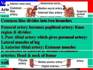
Emboli • Causes acute ischemia/ medical emergency • Most non-atherosclerotic emboli come from the heart • Entire lower extremity can be involved, most occlude lower leg, foot or toes. • Heparin, thrombolytic therapy, embolectomy • Common
Pseudoaneurysm • Mostly traumatic • Infection is most serious complication • Can be painful • Rarely causes ischemia • Occurs more in lower extremity than upper
Popliteal Artery entrapment • Popliteal artery compression by medial head of gastrocnemius muscle. • Young patients • With exercise the gastrocnemius muscle contraction compresses artery. • Uncommon
Trauma • Dissection, thrombosing, Arterial/venous fistulae. • Acute ischemia • Can happen to upper or lower ext. • Fairly common
Arterial-Venous Fistulas • Can be surgically created in lower ext for hemodyalisis. Occurs less frequently than in upper. Complications: aneurysm, pseudoaneurysm, infection, graft occlusion. • Fairly common (more often in arms) • Traumatic: • Artery and venous connection due to trauma • Infection is most serious complication over ischemia • Can be painful
Raynaud’s Disease • Affects upper extremities more significantly than lower. • Vasospasm without underlying occlusive or systemic cause. • Uncommon in lower
Arteritis • All types of arteritis affects upper extremities much more frequently than lower. (Takayasu’s, Giant Cell Arteritis, Polyarteritis or periarteritis. • Uncommon to see these listed as cause for lower extremity ischemia, but is possible.
Buerger’s Disease • Thromboangiitis Obliterans: described as rarely a cause, and accounting for less than 1% of lower extremity vascular disease. • Young, male, smokers, digit ischemia • Sudden onset • Claudication of foot and arch rather than legs. • Associated superficial thrombophlebitis • Less likely to cause lower ext ischemia than upper extremity ischemia.
Advential Cyst • Cyst of advential layer of arterial wall, causing stenosis or occlusion by thrombosing. • Can cause claudication • Can be surgically drained or bypassed. • Can reoccur • Uncommon
Hypercoagulability • Heparin induced thrombosis • Antithrombin III deficiency • Abnormal fibrinolytic system • Abnormal platelet aggregation • Uncommon
Hematologic disease • Polycythemia Vera • Thrombocytosis • Dysproteinemias. • Sudden onset, usually affects digits • Hematology consult for therapy • Uncommon
Summary of Lower extremity arterial complications • Common • Emboli • Pseudoaneurysm (Lower ext arterial injury, does not usually cause ischemia)
Summary lower cont. • Fairly common • Popliteal artery entrapment • Trauma • Arterial – venous fistula
Summary Lower ext arterial • Uncommon in Lower ext ischemia • Raynaud’s disease or phenomena • Arteritis • Buerger’S Disease • Advential cyst • Hypercoagulability • Hematologic disease
References Vas Phy 3 • Slides 1,2,3,4,5 Taber’s cyclopedic Medical Dictionary, Davis 1985 • Slide 5 Intro to Vascular Ultrasonography, Zwiebel, Saunders, 2000, Pg258 • Slides 6,7,8 Intro to Vascular Ultrasonography, Zwieber, pgs 259-260. & Handbook of Patient Care in Vascular Disease, 4th Ed., Hallett, Brewster, Rasmussen pgs 238-247
References Vas Phy 3 cont • Slide 9 Intro to Vascular Ultrasonography, Zwiebel, 2000, pg 259. & Cardiology Clinics, PVD in The Elderly, Breslin Ed., August 1991, pgs 559-560. • Slides 10,11 Intro to Vascular Ultrasonography, Zwiebel, 2000, pgs 259-261. & Vascular Diagnosis 4th Ed, Bernstein, Mosby, 1993. • Slide 12 Vascular Diagnosis 4th Ed., Bernstein, Mosby, 1993 pg 631
Refer Vas Phy 3 cont. • Slides 13,14,15 Cardiology Clinics, August 1991 pgs547-552. & Vascular Diagnosis, Bernstein, pg 631 • Slides 16,17 Intro to Vascular Ultrasonography, Zweibel, 2000 Pg 260. • Slide 20 Handbook of Patient Care in Vascular Disease 4th, Hallett, pg37. • Slide 21 Cardiology Clinics, August 1991,pgs 501
Reference Vas Phy 3 cont. • Slide 22 Intro to Vascular Ultrasonography, Zweibel, 2000, pg205. & Cardiology Clinics, August 1991 pgs 559-560 • Slide 23,26 Cardiology Clinics, August 1991, pg501. • Slide 27 Cardiology Clinics, August 1991, pgs 501-502/ • Slide 29 Cardiology Clinics, August 1991, 497-513.