Download
1 / 43
570 likes | 2.98k Views
Osteomyelitis and Septic arthritis. นพ.สุรพงษ์ ลีโทชวลิต 8 กันยายน 2551 . Complication . Sepsis, Toxic shock disability and deformity. Classification . Age group Neonate Childhood Adolescent Duration of symptom Acute Sub-acute Chronic. Classification . Route of infection

E N D
Osteomyelitis and Septic arthritis นพ.สุรพงษ์ ลีโทชวลิต 8 กันยายน 2551
Complication • Sepsis, Toxic shock • disability and deformity
Classification • Age group • Neonate • Childhood • Adolescent • Duration of symptom • Acute • Sub-acute • Chronic
Classification • Route of infection • Hematogenous system** • Direct inoculation :Open Fx, operation, skin puncture • Soft tissue infection • Causative organisms • Pyogenic organisms** • granulomatous
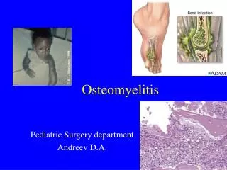
Natural history and Pathogenesis of Acute hematogenous osteomyelitis • Almost at “metaphysis” • lower extremities > upper extremities 5 เท่า โดยเฉพาะที่ distal femur และ proximal tibia • metaphysis (no phagocytosis cell) ≠ diaphysis (diaphysis = reticuloendothelial tissue + phagocytosis cells)
Source of infection • Blood circulation : • infection in Oral, Throat, Ear, Gastrointestinal tract, Urinary tract, Skin and soft tissue • Trauma (30-50%) • หกล้ม กระแทก • Minor trauma Caution : in infant < 18 month มักเกิด septic arthritis ร่วมกับ Osteomyelitis
Source of Infection Pathogenesis Blood stream Metaphysis Venousstasis Bacterialcolonization
Inflammation: acute osteomyelitis • First 24 hours • Vascular congestion • Polymorphonuclear leukocyte infiltration • Exudation
Inflammation: acute osteomyelitis • 2-3 day No treat with antibiotic • Intraosseus pressure intense pain intravascular thrombosis ischemia เด็กจะร้องปวดมาก
Suppuration • 4-5 days • Pus formation • Subperiosteal abscess via Volkmann canals • Pus spreading • epiphysis • joint • medullary cavity • soft tissue
Necrosis • Bone death by the end of a week • Bone destruction ← toxin ← ischemia • Epiphyseal plate injury • Sequestrum formation • small removed by macrophage,osteoclast. • large remained
New bone formation • By the end of 2nd week (10 – 14 days) • Involucrum (new bone formation from deep layer of periosteum ) surround infected tissue. • If infection persist- pus discharge through sinus to skin surface Chronic osteomyelitis
Femoral head and neck ( hip ) Humeral head ( shoulder ) lateral side of distal tibia ( ankle joint ) radial head and neck ( elbow joint ) joint capsule of 4 metaphysis cause of osteomyelitis
Signs and Symptoms in infant • Drowsy • Irritable • Fails to thrive • history of birth difficulties • History of umbilical artery catheterization • Metaphyseal tenderness and resistance to joint movement
Signs and Symptoms in child • Severe pain • Malaise • Fever • Toxemia • History of recent infection • Local inflammation pus escape from bone • Lymphadenopathy
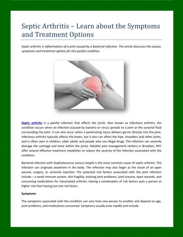
Natural History and Pathogenesis of Septic arthritis • Septic arthritis พบบ่อยที่ข้อใหญ่ๆ มากกว่าข้อเล็ก เป็นsingle joint painยกเว้นใน neonate ที่พบมากกว่า 1 ข้อ • 35 - 50 % พบที่ HIP • 20 – 25 % พบที่ Knee • ที่เหลือเป็น shoulder , ankle , elbow etc
Source of infection • Same as hematogenous osteomyelitis : • Blood circulation • Post operation • Skin and tissue infection etc.
Complication • Synovial fluid = good culture media • Within 8 hr. loss of glycosaminoglycan Wear and tear synovitis Cartilage destruction in 5 day
Signs and symptoms in newborn • Clinical of septicemia : fever (36 - 74 %) irritable, refuses to feed, rapid pulse • Joint swelling • Tenderness and resistance to movement of the joint • Look for umbilical infection
Signs and symptoms in children • acute pain in single joint ข้อใหญ่ๆ : hip. • Pseudoparesis. • Swelling and inflammation of the joint. • Child looks ill. • Limit movement of the joint. • Look for a source of infection : toe, boil, otitis media
Diff. diagnosis • Toxic synovitis เจ็บมากเป็นบาง direction ของการเคลื่อนไหว แค่ทำ Rolling test เบาๆ ก็เจ็บมาก flex เพิ่ม , extend หรือ rotate ไปทางไหนก็เจ็บไปหมด • Toxic synovitis ( transient synovitis ) มักจะเดินได้แต่กะเผลก Rolling test ไม่เจ็บ เจ็บเฉพาะทำ internal ratation • Juvenile rheumatoid arthritis จะเห็นข้อบวม แดง ร้อน ไม่ค่อยเจ็บมากเท่าไร ยังขยับข้อได้ดีพอควร ไม่ look sick กินได้ เล่นดีอยู่
Diff. diagnosis • Cellulitis จะเจ็บที่ผิวหนังภายนอก มีบวม แดง ร้อน ให้เห็นชัด แต่ขยับเคลื่อนไหวข้อไม่เจ็บ • Pyomyositis เด็กจะเจ็บตรงกล้ามเนื้อที่เป็น กล้ามเนื้อที่พบบ่อยคือกล้ามเนื้อต้นขา ทำให้เดินไม่ค่อยไหว มีไข้ ต้นขาบวมลึกๆ อุ่นๆ • Psoas abscess เจ็บที่ขาหนีบและท้องน้อย เดินได้แต่ต้องงอข้อสะโพกไว้ตลอด
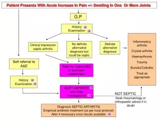
Investigation after admit • CBC • U/A • ESR • aspirateข้อ หรือ bone ( metaphysis ) • Gram stain of synovial fluid • C/S • Plain film
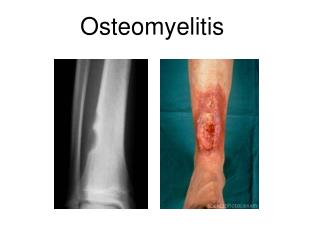
Investigation : Plain film • มักจะเปลี่ยนแปลงหลังจากการติดเชื้อนานกว่า 10 วัน • เริ่มจาก periosteal new bone formationrarefaction, area of lytic and sclerotic lesion, sequestrum and involucrum. • ควรเริ่มให้การรักษาทันทีก่อนจะเห็นการเปลี่ยนแปลงในภาพถ่าย X-ray
Investigation: ultrasound Ultrasound: ช่วยบอกว่ามี effusion ในข้อได้ ในบางครั้งก็บอกได้ว่า เป็น multiecchoic ซึ่งอาจจะหมายถึง Pus and fibrin มีการใช้ ultrasound ในการช่วยบอกตำแหน่ง ของการเจาะดูด และ follow up ดูว่า effusion ลดลงหรือไม่ หลังให้การรักษา
Investigation: MRI • ข้อบ่งชี้ : infection ที่ spine หรือ กรณีสงสัยว่าจะเป็นโรคอย่างอื่นมากกว่า เช่น tumor MRI • ใน Osteomyelitis • จะเห็นมีการลดลงของ marrow signal intensity ใน T1 wieghted images • หรือ การเพิ่มขึ้นของ marrow signal intensity ใน T2 weighted images สาเหตุจาก marrow fat ถูกแทนที่ด้วย inflamatory cells และedema
Investigation: CT scan • เห็น extent ของ bone destruction ได้ดี • ช่วยในการวางแผนการผ่าตัด exposure โดยเฉพาะ Osteomyelitis ของ spine หรือ pelvis
Investigation : Bone Scan • ใช้ในกรณีที่ไม่สามารถ locate lesion ใน early stage หรือมีหลายจุด(foci) • 99m TC-HDP - sensitive - not specific • แบ่งเป็น 2 phases • vascular phase warm (hot) uptake ขึ้นกับเลือดที่มาpool ตำแหน่งที่มี swelling & inflamation มาก • ต่อมาเป็น osseous phase ซึ่ง warm (hot) uptake จะเห็นตรง bone ที่มี lesion การเจาะข้อหรือเจาะ bone ไม่ได้ทำให้ผล bone scan เปลี่ยนแปลง
Investigation : Aspiration • confirm diagnosis • ต้องเจาะก่อนให้ Antibiotic • smear for cell and organism • culture and sensitivity test
Synovial fluid cells count analysis • Early case :WBC 25,000 – 50, 000 / μL • Late case : WBC count >50000 / μL (Juvenile rheumatoid arthritis พบได้เช่นกัน) • > 3 day พบ PUS ควรจะผ่าตัดล้างข้อทันที เพื่อเอา debris tissue, fibrin exudate และenzymes ย่อยสลาย cartilage ออก + ใส่ close suction drainageไว้
Antibiotic treatment • Cloxacillin dose ที่ให้คือ 150 – 200 mg/kg/day IV โดยแบ่งให้ทุก 4 – 6 ชั่วโมง สูงสุดไม่เกิน 12 gm./day (Neonate ลด dose เป็น 50 mg/kg/day) • Gentamicin ใช้กับ Gram negative rod ได้ผลดี dose ที่ให้ 2.5 mg/kg/8 to 24 hr. ปรับตามค่า Cr. • Gentamicin มักจะให้ร่วมกับ Cloxacillin ในระยะแรกที่ยังไม่รู้เชื้อ และถ้าจำเป็นต้องให้นานกว่า 1 สัปดาห์ ต้องระวัง Nephrotoxicity เจาะ BUN, Cr ทุก 3-4 วัน ถ้าสูงขึ้นต้องหยุดยา
Antibiotic treatment • Cefotaxime 100 – 200 mg/kg/day IV แบ่งให้ทุก 6 – 8 ชั่วโมง (Neonate ลด dose เป็น 50mg/kg/day แบ่งให้ทุก 8 – 12 ชั่วโมง) หรือ Ceftriaxone 50 – 100 mg/kg/day แบ่งให้ทุก 12 ชั่วโมง ใช้ได้ผลดีกับ H.influenza , Salmonella, Neiserria gonorrhea • อาจใช้ Cefazolin 100 – 150 mg/kg/day แบ่งให้ทุก 8 ชั่วโมง แทน Cloxacillin ได้ • กรณีแพ้ยากลุ่ม penicillin ให้ใช้ Clindamycin 30 – 40 mg/kg/day
Criteria Switch antibioticIV to Oral • Compliance of patient • Clinical sign and symptom: Fever <38 C (>72 hr.) , reduce pain , • CRP lower to normal • Switch to High dose oral antibiotic : Cloxacillin , cefazolin Cloxacillin , Cephalexin 100 – 150 mg/kg/d divided qid maximum dosage : 4 g/d
Subacute osteomyelitis • อาการไข้ไม่ชัดเจน • เจ็บกระดูกพอทนได้ • ถ้าเป็นที่ขาจะเดินได้แต่กะเผลก • สภาพทั่วไปของเด็กดีกว่า • ESR สูงไม่มาก CRPไม่สูง • Hemoculture ไม่ขึ้นเชื้อ Plain • film จะเห็น lytic lesion with or without sclerotic border มักพบที่ metaphysis
Chronic osteomyelitis • เกิดจากการ delay diagnosis and treatment ของ Acute osteomyelitis • ทำให้มี sequestrum,involucrum และ chronic sinus drainage โดยทั่วไป sequestrum จะเห็นได้ชัดเจนใน plain film การรักษา • ผ่าตัด debridement, sequestrectomy, saucerization • ร่วมกับการให้ยาปฏิชีวนะ IV pre OP และ post OP ต่ออีก 7 วันเปลี่ยนเป็นยากิน high dose ต่อจนครบ 6 สัปดาห์ หรือจน ESR < 25 สิ่งสำคัญคือต้อง remove sequestrum ซึ่งเป็น dead bone ไม่มี blood supply ทำให้ยาและphagocytic cells ไม่สามารถเข้าไปกำจัดเชื้อโรคที่หลบซ่อนอยู่ได้
ขอขอบคุณ • ภาพจาก presentation ของ ผศ.นพ.ยงศักดิ์ หวังรุ่งทรัพย์ ภาควิชาออร์โธปิดิกส์ จุฬาลงกรณ์มหาวิทยาลัย