Download
1 / 55
550 likes | 1.35k Views
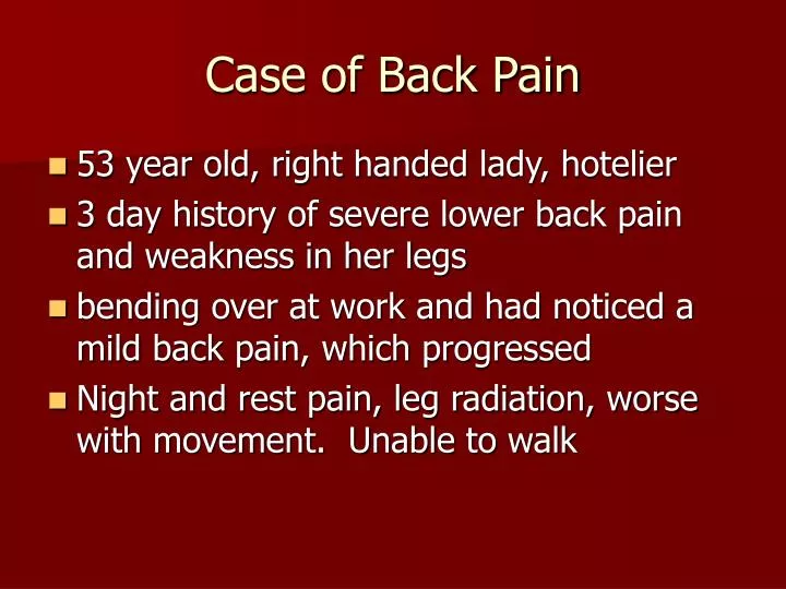
Case of Back Pain. 53 year old, right handed lady, hotelier 3 day history of severe lower back pain and weakness in her legs bending over at work and had noticed a mild back pain, which progressed Night and rest pain, leg radiation, worse with movement. Unable to walk. Case of Back Pain.

E N D
Case of Back Pain • 53 year old, right handed lady, hotelier • 3 day history of severe lower back pain and weakness in her legs • bending over at work and had noticed a mild back pain, which progressed • Night and rest pain, leg radiation, worse with movement. Unable to walk
Case of Back Pain • Sep 05Haematologists shoulder pains, lymphadenopathy and rash, fatigue, 7 kg weight loss in 6 months • l-node < 1cm ALP 210 Rheum referral • Subsequently admitted • Ex In pain restricted spine ? leg weakness and altered sensation feet
Case of Back Pain • ALP 320, ALT 89 CRP 96 XR normal • MRI spine normal • Symptoms progressed • Tingling in upper limbs, noted to have reduced reflexes
Case of Back Pain • CSF protein 2.55 g • ?Guillan-Barre • Transferred to neurology • IV Ig, Rehab, FVC, vitals monitoring • Campylobacter IgG and IgA 160 • EBV +ve
GB syndrome • Post-infective acute inflammatory demyelinating polyneuropathy • 1-3 weeks post viral • Distal numbness and weakness – evolves over days to weeks ascending • Back and leg pain can be a feature • 20% severe with autonomic and respiratory complications • Weakness, areflexia, sensory loss
GB syndrome • Rare – ocular and ataxia – Miller-Fisher syndrome • NCS: slowing of conduction or block • CSF: 1-3g/l • IV Ig, supportive, ventilation, plasmapharesis, rehab
BACK PAIN Jaya Ravindran Rheumatologist
Causes • Simple mechanical eg ligamentous strain • Degenerative disease with/without neural, cord or canal compromise • Metabolic – osteoporosis, Pagets • Inflammatory – Ankylosing spondylitis • Infective – bacterial and TB • Neoplastic • Others, (trauma,congenital) • Visceral
Red flags • Age <20 or >50 with back pain for the 1st time • Thoracic pain >50 yrs • Pain following a violent injury/trauma • Unremitting, progressive pain
Red flags • Past or current history of cancer • On Steroids or immunosuppressants • Drug abuser or +ve HIV • Systemic symptoms - fever, appetitie and weight loss, malaise
Red flags • Bilateral leg radiation, sensory/motor/sphincter symptoms • Pain predominantly at night
Inflammatory flags • Morning stiffness and pain >30 mins -1 hr • Better with activity • Peripheral joint involvement • Anterior uveitis • Psoriasis • Inflammatory bowel disease • Recent GI or GU infection • Family history
Myotomes • C5 Deltoid, biceps (biceps jerk) • C6 Wrist extensors, biceps (biceps, brachioradialis jerk) • C7 Wrist flexors, finger extensors, triceps (triceps jerk) • C8 Finger flexor, thumb extensors (triceps jerk) • T1 finger abductors
Myotomes • L2 Hip flexion • L3 Knee extension (knee jerk) • L4 Knee extension, ankle dorsiflexion (knee jerk) • L5 toe dorsiflexion • S1 foot plantar flexion, eversion
Examination • LOOK – deformity, muscle wasting, kyphosis, scoliosis • LOOK – normal cervical lordosis, thoracic kyphosis, lumbar lordosis • FEEL – spinal processes and sacroiliac joints
Examination • MOVE – Lumbar flexion • Schober’s test – marks at “dimples of Venus” and 10 cm above. Measure at maximal flexion – usually 5 cm • MOVE – Lumbar lateral flexion • MOVE – Cervical flexion/extension, lateral rotation and flexion, thoracic rotation
Examination • Sciatic stretch (patient supine) - Straight leg raise and dorsiflexion of foot - pain in calf and posterior thigh between 30-70o – low lumbar (L5/S1) lesion or sciatic irritation • Femoral stretch (patient prone) – knee is flexed and then hip extended – pain in anterior thigh – high lumbar (L2-L4) lesion
Imaging • XR – tumour, fracture, infection, inflammation • Bone scan – increased turnover eg infection, metastatic disease, fractures, Pagets • MRI – soft tissue, discs, facet joint, nerve roots, cord, inflammation
Degenerative disease and sciatica • Very common • Facet joint OA, disc disease, osteophyte • Mechanical back pain • Sciatica – most resolve NB persistent, neurology, bilateral, red flags • Analgesia, PT, pain clinics
Malignancy • Unremittting, progressive and night pain • Systemic symtoms • Past hx Ca • Breast, bronchus, thyroid, kidney, prostate and myeloma/plasmacytoma • Osteolytic (prostate osteoblastic) • XR can be normal in early stages – further imaging if suspicion high • Predilection for vertebral body and pedicles
Infection • discitis, osteomyelitis, and epidural abscess. • hematogenously spread • most often Staphylococcus aureus. • Gram-negative rods in postoperative or immunocompromised patients • normal skin flora is less commonly isolated in postoperative patients. • Postoperative patients develop symptoms 2 to 4 weeks after surgery after an initial improvement in pain.
Infection • Pseudomonas organisms in intravenous drug users. • Mycobacterium tuberculosis in developing nations and immigrant population. Fungal infections are rare. • Only one third have fever and 3% to 15% present with neurologic deficit. • Infections typically involve the intervertebral disc and vertebral body endplate
Infection • Radiographic changes at 2 to 4 weeks • bone scan can be positive as early as 2 days 75% specific. • MRI appearance is decreased T1- and increased T2-weighted signal in the infected disk. Enhancement after gadolinium
Infection • Conservative treatment of antibiotics, rigid bracing to prevent deformity and control pain • Surgery : neurologic deficit, presence of abscess, extensive bone loss with kyphosis and instability, failure of blood work and biopsy to isolate any organism, excision of a sinus tract, or no response to conservative treatment.
Osteoporosis - risks • History of low trauma # - colles, NOF, vertebral, sacral or pelvic insufficiency • Steroids • Maternal history of NOF # • Gonadal hormone deficiency • Ca deficiency • Prolonged immobility • Low BMI • Alcohol and smoking
Osteoporosis • Bisphosphonates • SERMs • Strontium • Teriparatide • Calcitonin • Lifestyle factors • Ca and Vit D
7-dehydrocholesterol sunlight cholecalciferol • (diet) • liver • 25-hydroxycholecalciferol • kidney 1-hydroxylase • 1,25-dihydroxycholecalciferol (-) • increased GI Ca2+ absorptionCa2+ • Bone resorption Thyroid • (-) • Parathyroid Gland PTH Renal Ca2+(-) Calcitonin • reabsorption
Spinal stenosis • Canal or foraminal narrowing with possible subsequent neural compression • Cause • Ligamanetum flavum hypertrophy, facet joint hypertrophy, vertebral body osteophytes, herniated disc • Rare: Pagets, AS, acromegaly
Spinal stenosis • Symptoms • Age - >50 • Dull aching pain in the lower back and legs • Exertional leg pain/weakness/numbness • Symptoms relieved leaning forward, sitting or lying • Examination • May be normal • Normal sensation and power • Reflexes normal or slightly reduced • Normal foot pulses
Spinal stenosis • Conservative – analgesics, NSAIDs, PT, epidural • Surgery – laminectomy (+arthrodesis)
Cauda Equina Syndrome • Back pain, lower limb weakness, saddle anaesthesia, sphincter disturbance, impotence • Causes – usually disc, rarely tumour, abscess, advanced AS • Diminished sensation L4 to S2 (sacral numbness), weakness ankle and plantar dorsiflexion, loss ankle jerks, urinary retention, loss anal tone • Urgent MRI and surgical decompression
Pagets • Pain, deformity • Skull, long bone, vertebra, pelvis, near hip • Neurologic compromise • Planned surgery • ?ALP 2X ULN • Rare: high output failure