Download
1 / 53
610 likes | 2.14k Views
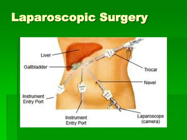
Risk of biliary tract injury during laparoscopic surgery. By Dr Fadhl Ali Almohtady Consultant Surgeon UST-Hospital 30--31 /5/2o12. INTRODUCTION. Open cholecystectomy was the standard practıce for treartment of symptomatıc gallbladder dısease untıl later 1980

E N D
Risk of biliary tract injury during laparoscopic surgery By Dr Fadhl Ali Almohtady Consultant Surgeon UST-Hospital 30--31 /5/2o12
INTRODUCTION Open cholecystectomy was the standard practıce for treartment of symptomatıc gallbladder dısease untıl later 1980 At present more than 90% of cholecystectomıes are performed by laparoscopy whıch become one of the commonest surgıcal procedure ın the world. Unfortunately the wıdespread used of laparoscopy has lead to a concurrence rıse ın the ıncıdence of major bıle duct ınjury(BDI)
Complications of Laparoscopic Cholecystectomy :A National Survey of 4,292 Hospitals and an Analysis of 77,604 Cases Deziel D J et al Chicago Illinois - Am J of Surg 165 January 1993 • 1.750 respondents • 1.2% laparotomy for treatment of complications • 0.6% mean rate of bile duct injury (exclusive of cystic duct), that will be lowered after performing > 100 LC • 50% of bile duct injury was recognized postoperatively, required anastomotic repair • 33 pts died, 18 of them due to operative injury • 0.14% bowel injuries • 0.25% vascular injuries Most lethal complications
Bılıary ınjury durıng cholecystectomy OC :has been associated wıth 0.2%-0.4% risk of BDI. ON THE OTHER HAND (LC): has been associated with 2.5 fold to 4 folds increase in the incidence of post operative BDI. in 1990 high rate of BDI ıs due to in part to learnıng curve effect . A surgeon had ı.7% chance of BDI ın the first case a 0.17%chance of BDI after the 50th case. However,most surgeon passed through the learning curve (steady state)
BILE DUCT INJURY (I) • Any injury to the bile duct during cholecystectomy is a dreaded complication. • Major bile duct injuries may require biliary-enteric reconstruction • Many patients, their consultants, and their lawyers believe these treatments result in a lifetime of disability (Maraca R.J et al : Arch Surge 2003, 137:889-894)
BILE DUCT INJURY (2) • The occurrence of an accidental bile duct injury strikes the patient and surgeons with great force, as neither is prepared for this complication • Often the surgeons is not immediately aware of disaster, and a delayed diagnosis adds further difficulty to the potentially disturbed relationship between doctor and patient. (Gouma DJ and Obertrop H : BJS 2002,89,385-386)
The Problem • LChas been associated with a higher incidence of IA bile duct injuries • LC—0.4 to 0.8% • Traditional OC—0.2-0.4% • Association: • Increased mortality and morbidity • Reduced long-term survival • Reduced quality of life • Between 34% and 49% of surgeons are expected to cause such an injury during their career. • Awareness and preventative methods are of clinical importance to surgeons.
Risk Factors and Mechanism • Risk Factors • Surgeon related rısk factors • Lack of experıence • Mısıdentıfıcatıon of bılıary anatomy • Intraoperatıve bleedıng • Over confıdant surgeon • Improper ıterpretatıon of ıoc • Improper lateral retractıon (insufficient or excess • Lack of conversıon ınto OC ın dıffıcult cases
Risk Factors…….cont • Patıent related rısk factors: • Age & sex • Anatomical variations (biliary and vasculature) • Severıty of dısease : Acute ,chronıc cholecystıtıs,empyema and mırızzı syndrome,….. • prevıous surgery wıth adhesıons. • Obesıty
Common Variant's of bile duct anatomy Lacey Clinic, Burlington, MA.1994
MANNER OF CONFLUENCE RIGHT SECTORAL DUCTS Blumgart LH. Surg Clin N Am. 1994.74.4
Risk Factors…….cont • Rısk factors ınherent to laparoscopıc approach; • 2-dimensional video monitor view, fixedviewpoint, etc .loss of depth perceptıon Lack of manual palpatıon Surgeon dependant to equıpment Blınd manıpulatıon the ınstruments
Mechanism of injury • Initially…Surgeon’s Learning Curve –Steady • Anatomical Misidentification: excision, incision, or transection of biliary anatomy • Injuries: common bile duct, common hepatic duct, right and left hepatic ducts, right hepatic artery, ducts draining hepatic segments • Anatomical variations (biliary and vasculature) • Electro cautery, thermal injury: stricture of CBD or hepatic ducts, bile leak • Mechanical trauma: stricture of the biliary ducts, bile leaks • Improper surgical approach
Misidentification injuries 2 main types ; • 1-CBD is mistaken for cystic duct so is clipped and divided. • 2-The segment of an aberrant right hepatic duct at the junction of cystic duct and CHD is mistaken for cystic duct
Classic Laparoscopic Injury --Mistaking the common bile duct for the cystic duct
Thermal Injuries • Inappropriate use of electro cautery near biliary ducts • May lead to stricture and/or bile leaks • Mechanical trauma can have similar effects Lahey Clinic, Burlington, MA.1994
CHD DRAINS FREELY IN TO THE PERITONEAL CAVITY Lacey Clinic, Burlington, MA 1994
CLASSIFICATION OF BDI There are many classıfıcatıon systems, Bismuth ,McMahon, Strasberg, Amesterdam academic medical center;s classificatıon, Stewart and so and so……..
CLASSIFICATION OF BDI…..cont PURPOSE to know the severıty of the ınjury. Communıcatıon purposes between doctors and centers Treatment purposes –modalıty of treatment
Bismuth's classification (1982)[ Type Criteria 1Low CHD stricture, with a length of the common hepatic duct stump of >2 cm 2Proximal CHD stricture-hepatic duct stump <2 cm 3 Hilar stricture, no residual CHD, but the hepatic ductal confluence is preserved 4Hilar stricture, with involvement of confluence and loss of communication between right and left hepatic duct 5Involvement of aberrant right sectorial hepatic duct alone orwith concomitant stricture of the CHD.
Bile Duct Injuries Bismuth classification of bile duct strictures Lacey Clinic, Burlington, MA.1994
McMAHON classıfıcatıon TYPE OF INJURY CRITERIA 1-MAJOR BDI 2-MINOR BDI 1-Laceratıon > 25% of bıle duct dıameter. 2-Trasectıon of CHDor CBD. 3-Development of post-operatıve stıcture. 1-Laceratıon of<25% of BD dıameter. 2-Laceratıon of cystıc-CBD junctıon(buttonhole tear)
Strasberg Classification • Type A Cystic duct leaks or leaks from small ducts in the liver bed • Type B Occlusion of a part of the biliary tree, almost invariably the • aberrant right hepatic ducts • Type C Transection without ligation of the aberrant right hepatic • ducts • Type D Lateral injuries to major bile ducts • Type E Subdivided as per Bismuth classification into E1 to E5
Strasberg Classification, cont’d • E: injury to main duct (Bismuth) • E1: Transection >2cm from confluence • E2: Transection <2cm from confluence • E3: Transection in hilum • E4: Separation of major ducts in hilum • E5: Type C plus injury in hilum
AMESTERDAM ACADEMIC MEDICAL CENTER 4 TYPES OF BDI CAN BE IDENTIFIED (MCMOHAN) TYPE A:cystıc duct leak or leakage from a berrant or perıpheral hepatıc radıcles. TYPE B:major bıle duct ınjury wıth or wıthout concomıtant bılıary strıcture. TYPE C:bıle duct stıcture wıthout bılıary leakage. TYPE D:complete transectıon of BD wıth or wıthout excısıon a part of the duct.
. Stewart-wayclassificationof BDI(2004) ] Class Criteria Ⅰ CBD mistaken for cystic duct, but recognized Cholangiogram incision in cystic duct extend Ⅱ Bleeding, poor visibility Multiple clips placed on CBD/CHD ⅢCBDmistaken for cystic duct, not recognized CBD, CHD, or right or left hepatic ducts transected and/or resected Ⅳ Right hepatic duct (or right sectorial duct) mistaken for cystic duct Right hepatic artery mistaken for cystic artery Right hepatic duct (or right sectorial duct) and right hepatic artery transected
BUT NON OFTHESE CLASSIFICATION SYSTEM IS UNIVERSALLY ACCEPTED ASEACH HASITS OWN LIMITATION
About 25% of BDI discovered ıntra-operatıvely. About 25% of BDI dıscovered after 24-48 hs post-operatıvely. And about 50% of BDI present weeks ,months or years post operatıvely
CLINICAL PRESENTATION • Many injuries are unrecognizedd at the time of the initial operation, and their presentation will vary • Those with associated bile leak will present early and often acutely ill from bile peritonitis or sub-hepatic abscess
CLINICAL PRESENTAION….CONT • Those with an injury but not leak, usually develop jaundice sometime after discharge from hospital, depending of the nature of the injury • Some injuries evolve slowly or cause partial obstruction • Stricture may involve principally the right or left hepatic duct or one of the right sectorial hepatic ducts
BILE LEAK IS RECOGNIZED EARLIER Presentation: • Acutely ill • Gut failure Warko karnadihardja- 2004
Intraoperative Detection • Only 25% of injures are recognized intraoperatively • If experienced, convert to Open Procedure and perform Cholangiography (determine extent of injury) • If not experienced, perform the cholangiogram laparoscopically with intent of referring patient (placement of drains) • Consult an experienced hepatobiliarysurgeon. Quicker the repair, the better the outcome!!!
Post-Operative Detection Plan • Controlling sepsis, establish biliary drainage, • Broad-spectrum antibiotics. • No need for urgent reconstruction of the biliary tree. • Reconstruction of the biliary tract is best performed electively after an interval of at least 6 to 8 weeks.
TIPS & TRICKS TO DIAGNOSE BILE DUCT INJURY History of unexplained fevers, pain, abnormal liver function test results, or pruritus Should prompt an investigation
TYPES OF IMAGING INVESTIGATION • Ultrasonography : • May reveal the : • ductal dilatation and or fluid collection(biloma • Of little value if bile ducts are decompressed
TYPES OF IMAGING INVESTIGATION (2) • Cholangiography • PTC is superior to ERCP • MRCP : Noninvasive, provides striking images of biliary tree • HIDA scan ; may show presence of active bile leak and general anatomic site of leakage.
MRC ; • Demonstrating dilatation or stenosis of the biliary tract; and stones in the bile duct remnant; the pancreas; and pancreatic duct; • However it doesn’t allow concomitant therapeutic measures.
ERCP; PTC • Can provide an exact anatomical diagnosis of bile duct leak. • Allowing for treatment of the leak by appropriate decompression of the biliary tract
TYPES OF IMAGING INVESTIGATION • Contrast-enhanced CT • The best initial study • May define level of injury, fluid collection or ascites • Reveal lobar atrophy • For vascular ınjury; CT –angıography, MR -angıography