Download
1 / 15
150 likes | 272 Views
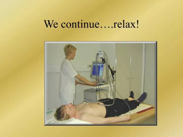
We continue….relax!. ST segment. ST segment changes. Terminology: Talking about specific lead (e.g. V2) depression is downward and elevation upward ….

E N D
ST segment changes Terminology: • Talking about specific lead (e.g. V2) depression is downward and elevation upward …. • Talking generally, we talk with respect to the closest leads(e.g. transmural damage causes ST segment elevation, while subepicardial damage causes ST segment depression) beware: When we talk generally about elevation, it can sometimes mean depression in the specific lead. E.g> Lateral Q- infarction will cause elevation of ST in V5, which can be, however, seen as depression in avR (the other side)
ST segment elevation Appears during ischemia, when subepicardial zone of myocardium is damaged – these cells depolarize with delay during the ST interval – we call this current of injury During transmural (all cardiac wall) damage, the epicardial zone prevails… Other causes: LV aneurysm, parasympathetic activation
ST segment depression Appears during ischemia, when subendocardial zone of myocardium is damaged – these cells depolarize with delay during the ST interval Appears in angina, and nontransmural (non –Q wave) MI Other causes: increased strain, digitalis medication digitalis
T wave • Physiologically: + : I, II, V3 až V6 • : avR +/- : III, avF, avL, V1, (V2) • The most variable part of ECG record. • Can become negative in increased intracranial pressure, pancreatitis, hypoK, hypoCa, non Q-wave MI • Can be physiologically negative in black people
HyperK Physiological: Repolarization goes from epicard to endocard (unlike depolarization)..T wave vector has similar direction like QRS vector “Tired” part of the myocardium repolarizes with delay. negative T: ”Tired” epicardium or whole wall. Positive but bigger pointing T: “Tired” endocardium Subendocardial ischemia
Non-Q wave MI Negative T (coronary) + depression of ST segment
Ischemie subepikardi- ální a transmurální myokarditis
U wave • Physiological: If appears, then small and with same polarity as T wave • Reflects later phases of ventricular repolarization (possibly repolarization of bundle of His) • Big and positive: hypoK – risk of arrhythmias • (big negative U can also appear with acute ischemia HypoK Milder Graver
Up to you to learn at home! Divided to bradyarrhythmias and tachyarrhythmias Tachyarrythmias can proceed to flutter and fibrillation Atrial fibrillation is the most common arrhythmia Ventricular fibrillation is the most common (80%) cause of sudden cardiac arrest Further Extrasystoles can appearSupraventricular x ventricular (How does look ES originating in left ventricle ?) Monoform ES x multiform Bigeminia, cuples, salves, phenomena R on T Arrhythmias
Sick sinus syndrom When upper centre stops the impulse generation is taken by subordinate centre It takes a while: Abram-Stokes’syncope AV block Bradyarrhythmias