Download
1 / 19
240 likes | 520 Views
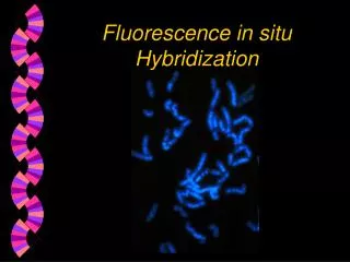
Immunolabeling & Fluorescent Microscopy. Presented by: Sumble Maha Khan ABE Workshop June 13 – 30, 2006. What is fluorescence?.

E N D
Immunolabeling & Fluorescent Microscopy Presented by: Sumble Maha Khan ABE Workshop June 13 – 30, 2006
What is fluorescence? Fluorescence is the bombardment of a pigment with high energy light (i.e. blue or UV), which in turn excites the pigment and emits light at a lower E and longer wavelength.
Goals of . . . Immunolabeling : To mark biological molecules or structures using antibody-antigen complexes. For example, the localization of proteins in cells. Fluorescent Microscopy : Is used to obtain a signal from the fluorescent probe on the antibody. For example, AlexaFluor 488.
Immunolabeling(using the two-step indirect method) • Sections of Arabidopsis provided on slides. • Pretreatment with glycine to keep autofluorescence to a minimum. • Series of washes & blockings performed to reduce the non-specific binding sites. • Overnight incubation with 1o antibody (anti-CNGC or anti-PDI-2). • Washes with TBST + 5% milk. • 1 hr incubation in dark with 2o antibody which has AlexaFluor 488 covalently attached to it. • Wash with TBS and place a wet coverslip with plain Vectashield mounting medium. • Seal and view under microscope.
Epi-fluorescence Microscope • Light source is a mercury arc lamp, has a broad band of excitation wavelengths. • Distributes single molecular species based on fluorescence emission properties. • Monitors precise location of intracellular components labeled with fluorophores. • Study factors such as pH, refractive index, ionic concentrations, membrane potential, solvent polarity.
Confocal Microscopy for high resolution images • Light source is a high-intensity monochromatic laser, which excites the fluorophore. • Minimizes background information so image does not degrade. • Controls depth of field (z-axis). • Spatial filtering eliminates the out-of-focus light or glare in specimens. • Collects serial optical sections of thick specimens, and constructs 3-D images using computer software.
PDI-2 & CNGC PDI-2 ~ protein disulfide isomerase It is one of the PDI family members. It facilitates protein folding by forming disulfide bonds. CNGC ~ cyclic nucleotide gated channel It regulates potassium ion transport in the cells, and it is located in the plasma membrane.
PDI-240x 1o ab ~ 1:10 anti-PDI-2 1o ab ~ 1:100 anti-PDI-2 2o ab ~ 1:100 AlexaFluor488 2o ab ~ 1:100 AlexaFluor 488
PDI-240x 1o ab ~ 1:10 anti-PDI-2 1o ab ~ 1:100 anti-PDI-2 2o ab ~ 1:10 AlexaFluor 488 2o ab ~ 1:100 AlexaFluor 488
PDI-240x glycine pretreatment no glycine pretreatment 1o ab ~ 1:10 anti-PDI-2 1o ab ~ 1:10 PDI-2 2o ab ~ 1:10 AlexaFluor 488 2o ab ~ 1:100 AlexaFluor 488
PDI-2 40x 1o ab ~ 1:100 anti-PDI-2 1o ab ~ none (control) 2o ab ~ 1:100 AlexaFluor 488 2o ab ~ 1:100 AlexaFluor 488
CNGC40x 1o ab ~ 1:10 anti-CNGC 1o ab ~ 1:100 anti-CNGC 2o ab ~ 1:100 AlexaFluor 488 2o ab ~ 1:100 AlexaFluor 488
CNGC 1o ab ~ 1:10 anti-CNGC 2o ab ~ 1:100 AlexaFluor 488
CNGC 1o ab ~ 1:10 anti-CNGC 1o ab ~ 1:10 anti-CNGC 2o ab ~ 1:100 AlexaFluor 488 2o ab ~ 1:100 AlexaFluor 488
CNGC40x 1o ab ~ 1:10 anti-CNGC 1o ab ~ 1:100 anti-CNGC 2o ab ~ 1:10 AlexaFluor 488 2o ab ~ 1:100 AlexaFluor 488
CNGC 40x 1o ab ~ 1:100 anti-CNGC 1o ab ~ none (neg. control) 2o ab ~ 1:100 AlexaFluor 488 2o ab ~ 1:10 AlexaFluor 488
Confocal Images Root10x Arabidopsis root ~ WT (neg. control) Arabidopsis root ~ GFP-2SC (pos. control)
Confocal Images Cotyledon 10x Arabidopsis cotyledon ~ WT Arabidopsis cotyledon ~ GFP-2SC (neg. control) (pos. control)