Download
1 / 102
1.07k likes | 1.26k Views
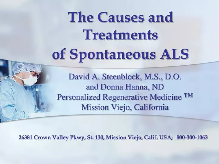
David A. Steenblock, M.S., D.O . and Donna Hanna, ND Personalized Regenerative Medicine TM Mission Viejo, California. The Causes and Treatments of Spontaneous ALS. 26381 Crown Valley Pkwy, St. 130, Mission Viejo, Calif, USA; 800-300-1063. Amyotrophic Lateral Sclerosis.

E N D
David A. Steenblock, M.S., D.O.and Donna Hanna, NDPersonalized Regenerative Medicine TMMission Viejo, California The Causes and Treatments of Spontaneous ALS 26381 Crown Valley Pkwy, St. 130, Mission Viejo, Calif, USA; 800-300-1063
Amyotrophic Lateral Sclerosis • ALS has been quite a mystery! • Within the spinal cord are certain neurons that control the muscular movements to a person’s extremities. • With ALS, these motor neurons become irritated, inflamed and over time slowly die, leaving the person totally paralyzed and leading to death within a few short years.
Glass CK, et al. Mechanisms Underlying Inflammation in Neurodegeneration, Cell 2010; 140: 918-934.
Amyotrophic Lateral Sclerosis My “wholistic” approach has been • to look at the whole body for clues to determine what the causes are and then • to find natural treatments that can remove the causes and correct the damage that has already been done by the disease process.
Primary Cause of ALS • Acute or chronic damage to spinal nerves.
Primary Cause of ALS 1. Damage to spinal nerves releases toxic cytokines including • Tumor Necrosis Factor alpha (TNF-α) • Gamma-interferon (γ-IFN) 2. The spinal nerve damage opens the spinal CSF-blood barrier which allows toxins to enter the CSF and spinal cord.
Primary Cause of ALS • Multiple forms/types of toxins that enter the CSF in ALS • Metals (mercury, lead, etc.) • Superoxide (O2-) • Cytokines • Nitric oxide (S-nitrosylation) • Intestinal bacteria • Yeast metabolites • Clostridial neurotoxins
Primary Cause of ALS • Lipoteichoic acid (from bacteria outer cell wall) • Endotoxins • MMP-9 • Chemicals, pesticides • Diesel fuel • Viruses • Prions, etc.
Primary Cause of ALS • Reactive oxygen species (ROS), endotoxins and inflammation in the intestinal tract can cause gut wall permeability (leaky gut syndrome), that allows the oxidative stress (ROS), endotoxins and inflammation to oxidize monocytes and T-regulatory cells. • Biofilm are also formed in the GI tract from E.coli, yeast, clostridia, etc. Menozzi A, Ossiprandi MC. Assessment of enteral bacteria. CurrProtocToxicol 2010, Chapter 21: Unit 21.3.
Primary Cause of ALS • Paneth cells (host defense cells in the GI tract mucosa) also secrete TNF-alpha. Paneth cells
Toxic microbials into the GI tract Destroying the intestinal wall Flowing into the bloodstream
ALS and Genetics • The general consensus is that only about 10% of ALS patients have the inherited genetic form of the disease (with SOD1 and/or TDP-43 gene mutations). • Most ALS cases are sporadic, having mutations in other genes. Wijesekera LC, Leigh PN. Amyotrophic lateral sclerosis. Orphanet J Rare Dis 2009; Feb 3; 4:3.
ALS and Genetics • Every ALS patient should have an overall genetic test which can be ordered from www.23andme.com (about $100.00). • The next step is taking that information to www.promethease.com and www.geneticgenie.com for gene-protein factors (about $15.00 each).
ALS and Genetics • Knowing a patient’s genetic mutations may help the physician determine the type of diet and treatments that will help promote better overall health.
Spinal Cord Injuries • Over the past few years, peri-spinal pain, impingement and avulsion (a forcible tearing away) of the spinal nerves, especially cervical 4-5 and C5-6, have been shown to produce increased oxidative stress through TNF-α and gamma interferon. C 4-5 C 5-6
Spinal Cord Injuries • An early article by Strickland and his research group (1996) found that ALS patients showed severe head, neck or back injury compared to their matched control group. Strickland D, et al. Physical activity, trauma, and ALS: a case-control study. Acta Neurol Scand 1996; 94(1):45-50.
Spinal Cord Injuries • Aminoguanidine has been used in rat studies to reduce permeability in the blood-spinal cord barrier. • In addition, the patient’s bone-marrow stem cells can be used to heal the injured spinal cord barrier so that reactive oxygen species (ROS) are stopped from flowing into the spinal cord. Fan ZK, et al. The effect of aminoguanidine on compression spinal cord injury in rats. Brain Res 2010, 25;1342:1-10.
Spinal Cord Injuries Degenerative conditions that have impinged the spinal arteries can lead to marginal oxygenation in the spinal cord. • Chronic stimulation of Vascular Endothelial Growth Factor (VEGF) leads to down-regulation of the VEGF gene. Schneeweis C, et al. Chronic CRP-exposure inhibits VEGF-induced endothelial cell migration. J AtherosclerThromb 2010; 17(2):203-12.
Spinal Cord Injuries • A reduction in the VEGF gene leads to a lack of blood vessel repair and chronic hypoxia. • Chronic hypoxia leads to an increase in cytokines, oxidative stress, and calcium influx into the motor neurons. A test for Nocturnal Oximetry is recommended for ALS patients.
Spinal Cord Injuries • If there is an intermittent oxygen deficiency, breathing oxygen at night will help to reduce free radical stress in the spine. de Carvalho M, et al. Percutaneous nocturnal oximetry in amyotrophic lateral sclerosis: periodic desaturation. Amyotroph Lateral Scler 2009; 10(3): 154-61.
Spinal Cord Injuries • An ALS patient should have a CT scan of his/her spine. • Generally, over the area that has a degenerative joint disease (DJD), the doctor can palpate this area for spinal nerve pain. • This area can then be treated with an injection of buffy coat from the patient’s bone marrow aspirate from the iliac crest.
Environmental Risk Factors The following is a list of environmental risk factors for ALS that may interact with genetic factors: • History of trauma to the brain and spinal cord, • A history of participation in varsity athletics, • A slim physique, • Strenuous physical activity, • Radiation, electrical shocks, • Cigarette smoking, • Heavy metal poisoning, and/or • Pesticide exposure.
Glass CK, et al. Mechanisms Underlying Inflammation in Neurodegeneration, Cell 2010; 140: 918-934.
TNF-α and The Motoneurons • Inflammatory TNF-alpha from oxidative stress plays an important role in the formation and acceleration of spinal cord damage and motor neuron degeneration. Xu L, et al. Oxidative stress in immune-mediated motoneuron destruction. Brain Res 2009; 1302: 225-32.
LPS and Motoneurons • An endotoxin is a toxic lipopolysaccharide substance in the outer membrane of gram-negative bacteria. Endotoxins = Lipopolysaccharides • Exposure to lipopolysaccharide (LPS) leads to the death of motor neurons in a dose- and time-dependent manner. Li B, et al. The NADPH oxidase is involved in lipopolysaccharide-mediated motor neuron injury. Brain Res 2008; 1226: 199-208.
The Microglia • Nuclear factor-kappa beta (NF-kB) is a master regulator of inflammation and is upregulated in the spinal cords of ALS patients. • Activation of NF-kB in microglia induces motor neuron death in vitro and in vivo. Frakes AE., et al. Microglia induce motor neuron death via the classical NF-kB pathway in amyotrophic lateral sclerosis. Neurons 2014; 81(5): 1009-23.
NF-κB, TNF-α and Glutamate • High levels of pro-inflammatory cytokines such as tumor necrosis factor-alpha (TNF- α) have been found in ALS patients. • TNF-α induces NF-κB activation that increases glutamate excitotoxicity of motoneurons. Tolosa L, et al. TNF-a potentiates glutamate-induced spinal cord motoneuron death via NF-κB. Mol Cell Neurosci 2011; 46(1): 176-86.
Microglia • Glutathione is an important detoxifying agent of the body. • Therefore, there must be plenty of this neuroprotector in the ALS patient’s body (precursor: acetyl-L-cysteine and intravenous glutathione).
MMP-9 and Motoneurons • Matrix metalloproteinases are responsible for the integrity of the basement membrane by extracellular matrix degradation. • MMP-9 is involved in the pathology of several neurological diseases, including ALS. • MMP-9 levels are increased by neurovascular damage and leads to motor neuron degeneration. Miyazaki K, et al. Disruption of neurovascular unit prior to motor neuron degeneration in amyotrophic lateral sclerosis. J Neurosci Res 2011; 89(5):718-28.
MMP-9 Activation MMP-9 is activated by: • Candida albicans, Helicobacter pylori • Epstein-Barr virus, rhinovirus, papillomavirus 8, herpesvirus 6, HIV, cytomegalovirus, hepatitis B • TNF α, NF-kappa B • Mutant SOD1 activates MMP-9 that leads to endoplasmic reticulum stress (ER stress) that triggers axonal death. Kaplan A et al. Neuronal matrix metalloproteinase-9 as a determinant of selective neurodegeneration. Neuron 2014; 81(2): 333-48.
GI Tract Inflammation Factors that promote inflammation in the GI tract include: • Methylmercury, lead, cadmium, etc. • Oxidative stress (ROS) • Tumor Necrosis Factor alpha (TNFα) • Gamma interferon (γ IFN) • Yeast, Candida, Clostridia • Matrix metallopeptidase 9 (MMP-9) • Misfolded superoxide dismutase (SOD) • Superoxide free radical
GI Tract Inflammation Doctor’s Data has the following tests. If one laboratory does the tests, it’s easier to compare results from one patient to the next as well as utilize for research purposes. • Comprehensive Digestive Stool Test plus ova and parasites (to determine what “bad” bugs are present in the gut). • Quantitative Urine Organic Acid Test (to verify and monitor the neurotoxins present). • Toxic element challenge test using DMPS to determine heavy metal poisons. • Additional tests as needed
Toxic Metals • There is an increased risk of ALS in people whose cerebrospinal fluid has higher levels of lead, mercury, cadmium, manganese, aluminum, cobalt, copper, zinc, vanadium or uranium. • These metals are directly toxic to neurons. Roos PM, et al. Metal concentrations in cerebrospinal fluid and blood plasma from patients with amyotrophic lateral sclerosis. Biol Trace Elem Res 2013; 151(2): 159-70. Junas-Morales R, et al. Environmental factors in ALS. Presse Med 2014; 43(5): 549-54.
Methylmercury Health Effects Nervous system deterioration Depletes glutathione peroxidase, binds to thiol groups Induces mitochondrial dysfunction, reduces ATP synthesis, increases DNA damage Hearing, speech, vision and gait impairment Causes involuntary muscle movements Disrupts endocrine gland function Causes difficulty with chewing and swallowing
Toxic Metals • A pattern of multiple toxic metals is often seen in the ALS cerebrospinal fluid. • Chelation therapy is recommended for patients with these neurotoxic metals. • Mercury needs to be removed either by DMPS IV. • Lead is removed by oral or IV calcium EDTA. • If both are high, use DMPS for the mercury first.
Yeast Biofilm • Yeast produce a microbial biofilm that promotes gut inflammation. • A part of the biofilm is gram negative bacteria which deposit fragments of their outer cell wall (endotoxins=lipopolysaccharides) into the gut wall.
Candida albicans • Candida albicans begins as a yeast and then grows into a fungus, developing hyphae (from the Greek word: “web” – of long, branching filament structures of a fungus).
Gut Inflammation • As the hyphae penetrate into the gut, tumor necrosis factor alpha (TNF α), gamma interferon (IFNγ), MMP-9 and other cytokines (small proteins involved in cell signaling) are produced in the gut wall, creating an acute phase inflammatory condition.
Clostridium difficile • In 2005, Dr.Longstreth and his research team published an article hypothesizing that a clostridial species causes ALS in susceptible individuals. • The bacteria would reside undetected in the gut and chronically produce a toxin that targets the motor neurons. Longstreth WT Jr, et al. Hypothesis: a motor neuron toxin produced by clostridial species residing in gut causes ALS. Med Hypoth 2005; 64(6): 1153-6.
Costridia Infection Clostridia infection in the GI tract
Clostridia Infection • Clostridium difficile kills 14,000 people a year in America alone. Other Clostridium species include: • Clostridium perfringens(Gangrene, Food poisoning) • Clostridium tetani(Tetanus) • Clostridium botulinum (Botulism) • Clostridium acetobutylicum • Clostridium haemolyticum • Clostridium novyi • Clostridium oedematiens • Heliobacteria are also in the Clostridia class. Bugs in the system.The Economist. 3 November 2012.
GI Tract Infections • Misfolded superoxide dismutase leads to an increase in the superoxide free radical. • This superoxide free radical can cause Colitis as well as Crohn’s Disease. • Lactobacillus plantarum increases SOD, reduces superoxide and reduces Colitis and Crohn’s Disease. Jang SE, et al. Lactobacillus plantarum CLP-0611 ameliorates colitis in mice by polarizing M1 to M2-like macrophages. IntImmuno-pharmacol 2014; 21(1): 186-92. Smits HH, et al. Selective probiotic bacteria induce IL-10-producing regulatory T cells. J Allergy ClinImmunol 2005; 115(6): 1260-7.