Download
1 / 95
1.2k likes | 2.73k Views
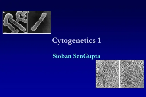
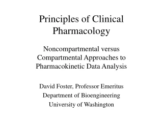
Principles of Clinical Cytogenetics. Clinical Cytogenetics is the study of chromosomes, their structure and their inheritance, as applied to the practice of medical genetics Chromosome disorders form a major category of genetic disease:

E N D
Clinical Cytogenetics is the study of chromosomes, their structure and their inheritance, as applied to the practice of medical genetics • Chromosome disorders form a major category of genetic disease: • Reproductive wastage, congenital malformation, mental retardation (MR), and pathogenesis of cancer. • Specific chr. abnormalities are responsible for 100’s of identifiable syndromes, collectively more common than single-gene disorders.
Prevalence of Cytogenetic Disorders ~1% of all live births ~ 2% of pregnancies in women older than 35 1/2 of all spontaneous first-trimester abortions
Indications for a karyotype • Problems of early growth and development: failure to thrive, developmental delay, dysmorphic faces, multiple malformations, short stature, ambiguous genitalia and MR. • Stillbirth and neonatal death • Fertility problems: couples with a history of infertility or multiple pregnancy loss, women with amenorrhea • Family history: a known/suspected chr. abnormality in a first degree relative • Neoplasia • Pregnancy in a woman of advanced age (>35 yrs)
A uniform system of chr. classification is internationally accepted for identification. • The pattern of bands on each chr is numbered on each arm. • By this numbering, location of any band, DNA sequences and genes within it, and its involvement in a chr. abnormality can be precisely described
Chromosome Identification • Culture (PB, fibroblasts, lymphobalstoid cell lines, Bone marrow, fetal cells) • Banding (G, Q, R) • Special procedures (C-banding, high resolution banding, fragile sites) • Molecular cytogenetics (e.g., FISH, SKY, CGH)
Ideogram, G-bands at metaphase, with about 400 bands/haploid karyotype.
Human chr’s are often classified by position of centromere into: metacentric, submetacentric, acrocentric. • Acrocentric (13,14,15,21,22) have small distinctive chromatin masses (satellites) attached to short arms by narrow stalks (secondary constrictions). • Stalks of these 5 pairs contain 100’s of copies of genes encoding rRNA as well as a variety of repetitive sequences.
Fluorescence in situ Hybridization • Confluence of genomic and cytogenetic approaches=molecular cytogenetics • Examine presence or absence of a particular DNA sequence or evaluate the number or organization of a chr. or chromosomal region.
FISH at metaphase and interphase with 3 different types of probes. F-VIII, α-satellite (chr. 17), painting probe (chr. X)
FISH • 2,3 and even 4-color applications to diagnose specific deletions, duplications, rearrangements in metaphase and interphase. • With specialized imaging procedure, 24 colors can be detected (SKY)
Chromosome and Genome Analysis by Use of Microarrays • Chromosome analysis can be performed at a genomic level by a variety of array-based methods using comparative genomic hybridization (CGH). • Assess the relative copy number of genomic DNA sequences in a comprehensive manner • Complements karyotyping and provides very sensitive, high resolution genome assessment. • Balanced translocations and rearrangements can not be resolved by array-based CGH.
Array CGH. Top: sample from a normal female. Bottom: sample from a male with trisomy 18.
Chromosome Abnormalities • Numerical • Structural • Balanced • Unbalanced • Stable vs. unstable
The most common type of clinically significant chr. abnormality is aneuploidy. Always associated with physical or mental maldevelopment or both. • Reciprocal translocations are also relatively common but usually have no phenotypic effect (risk of abnormal offspring) • Chr. abnormalities are described by a standard set of abbreviations and nomenclature and technology used (e.g., FISH or microarray)
The phenotypic consequences of a chr. abnormality depend on its specific nature, resulting imbalance of involved genome parts, specific genes involved, and likelihood of its transmission. • A number of general principles that should be kept in mind:
Unbalanced karyotypes in liveborns: General guidelines for counseling Monosomies are more deleterious than trisomies • Complete monosomies are generally not viable except for monosomy X • Complete trisomies are viable for chr. 13,18,21,X,Y. Phenotype in partial aneusomies depends on: • Size of unbalanced segment • Imbalance monosomic or trisomic, and • Region of genome and genes involved In a mosaic karyotype, “all bets are off” Rings give a phenotype specific to genome region involved, but are commonly mosaic. Inversions • Pericentric: risk of birth defects in offspring increases with size of inversion • Paracentric: very low risk of abnormal phenotype
Abnormalities of Chromosome Number • Heteroploid: any chromosome number other than 46 • Euploid: exact multiple of haploid • Aneuploid: any chromosome number other than euploid
Polyploidy-the presence of one or more extra complete sets of chromosomes in a cell Remember that diploidy (2N) is normal for human somatic cells and haploidy (N) is normal for germ cells Triploidy (3N) 69,XXX 69,XXY 69,XYY - Seen in fetuses, and lethal early in life - Observed in 1% to 3% of recognized conceptions - Usually caused by dispermy. Failure of one of the two meiotic divisions (diploid egg or sperm) may also occur. • Phenotype of triploid karyotype depends on source of extra chr. set: • Paternal abnormal placenta (partial hydatidiform moles), • Maternal spontaneously aborted earlier in pregnancy Tetraploidy (4N) 92,XXXX 92,XXYY - Much rarer than triploidy • Seen in fetuses, and lethal early in life • Absence of XXXY or XYYY suggests failure of completion of an early cleavage division of zygote
Aneuploidy • Cells that do not contain a multiple of 23 chromosomes (n) – there are missing or extra chromosomes • Most common and clinically significant chr disorder, present in at least 5% of all clinically recognized pregnancies • Trisomy: presence of three copies of a chromosome. Monosomy (less often): presence of only one copy of a chromosome. Both can have severe phenotypic consequences.
Most common type of trisomy in liveborns is trisomy 21. 47,XX or XY, +21: the constitution seen in 95% of Down syndrome. • Other trisomies observed in liveborns include trisomy 13, 18. • Notable that 13,18,21 are with low number of genes. • Monosomy for entire chr is almost always lethal. An important exception is X (Turner syndrome). • Aneuploidy is generally caused by chromosome nondisjunction • Premature separation of sister chromatids in M-I instead of M-II (another mechanism)
Karyotype of fetal cells with trisomy 21 (Down syndrome) Diagnosis: 47,XX, +21
Consequences of non-disjunction during meiosis I and II are different. • Non-disjunction has been associated with aberrations in frequency or placement, or both, of recombination events in meiosis-I. Too few (or even no) recombinations, or too close to centromere or telomere favor non-disjunction.
Classic nondisjunction: failure of chr’s either to pair or to recombine properly, or both. • Another mechanism involves premature separation of sister chromatids in meiosis I instead of II. Separated chromatids may by chance segregate to oocyte or to polar body unbalanced gamete.
More complicated forms of multiple aneuploidy • A gamete has an extra representative of more than one chr. • Nondisjunction can occur at two successive meiotic divisions, or by chance in both male and female gametes • Extremely rare except for sex chr’s. • Nondisjunction can occur in mitotic division after zygote formation. If early clinically significant mosaicism. E.g., in some malignant cell lines and some cell cultures
Probes: yellow/white for chr. 18; red for X; green for Y. Left: normal sperm cells Middle: 24,XX sperm Right: 24,XY sperm
An important diagnostic tool, especially prenatally, interphase multicolor FISH to evaluate 13,18,21,X,Y aneuploidy. Multicolor FISH analysis of interphase amniotic fluid cells
Structural Abnormalities • Breakage and reconstitution in an abnormal combination • Less common than aneuploidy • Present in about 1/375 newborns. • Chr rearrangements can occur spontaneously at low freq. & may be induced by clastogens, e.g., IR, some viral infections, and many chemicals. • Like numerical abnormalities may be present in all cells or in mosaic form • Balanced - no net gain/loss of chromosomal material • Unbalanced - gain/loss of chromosomal material • Stable: passing through meiotic & mitotic divisions unaltered • To be stable, a rearranged chr must have a functional centromere and two telomeres.
Unbalanced Rearrangements • The phenotype is likely abnormal. Any change that disturbs normal balance of functional genes abnormal development. • Deletions: lead to partial monosomy • Duplications: lead to partial trisomy • Large deletions/duplications (at least a few million bp) detected by karyotyping. • Smaller deletions/duplications requires FISH or microarray CGH analysis. Two-color FISH of a case with DiGeorge syndrome (deletion of 22q11.2).
Array CGH. A: partial duplication of 12p in a patient with an apparently normal routine karyotype and symptoms of Pallister-Killian syndrome. B: terminal deletion in 1p in a patient with mental retardation. C: de novo deletion in 7q22 in a patient with a complex abnormal phenotype
An important class involves submicroscopic changes of a telomere region in patients with idiopathic MR. small deletions, duplications & translocations have been detected in several percent of such patients • Targeted cytogenetic or genomic analysis of telomeric and subtelomeric regions by FISH or array CGH may be indicated in unexplained MR (important for counseling) A cryptic translocation in a developmentally delayed proband. Probes for telomere of chr. 3p (red) & chr. 11q (green). An unbalanced translocation b/w 3p and 11q.
Deletions • Caused by a break in a chromosome with a resultant loss of acentric segment/ unequal crossing over/ abnormal segregation from a balanced (translocation/inversion) abnormality • A carrier is monosomic for lost segment • Haploinsufficiency for those lost genes • Deletion may be terminal or interstitial • Clinical consequences depend upon size of deleted segment and number and function of lost genes
Analysis: • large deletions – visible cytogenetic changes (~ 1 in 7000 live births) • small deletions – high resolution banding/ Southern Blot/ Exon specific PCR/ FISH with targeted probes/ array CGH • Numerous deletions have been identified in dysmorphic patients & prenatal diagnosis. Knowledge of functional genes lost & their relation to phenotype.
Duplications • Originate by unequal crossing over or by abnormal segregation from meiosis in a carrier of a translocation or inversion. • Often lead to some phenotypic abnormalities • E.g., duplication of all or a portion of 12p leads to Pallister-Killian syndrome: characteristic craniofacial features, MR, and other birth defects likely related to trisomy or tetrasomy for specific genes in duplicated region
Marker and Ring Chromosomes • Marker chromosomes: very small, unidentified chromosomes, frequently mosaic. • Usually extra to the normal chr. complement; Supernumerary chromosomes or Extra Structurally Abnormal Chr. (ESACs) • Tiny marker chromosomes consist of little more than centromeric heterochromatin • Precise identification requires various FISH probes (SKY) • Larger markers contain some material from one/both arms imbalance for genes present
Prenatal frequency of de novo marker chr. ~1/2500 • Risk of fetal abnormality can range from very low to as high as 100% depending on marker origin • A relatively high proportion results from chr.15 and from sex chr’s. specific syndromes are associated with bisatellited chr.15 derived markers and with centric portion of X. • Neocentromeres: contained in a subclass of marker chr.; small fragments of chr. arms that somehow acquired centromere activity
Ring Chromosomes • Marker chr. that lack telomeric sequences • Deletion occurs at both tips of a chr. followed by a joining of the “sticky” chromosome ends • Rare, but have been detected for every chr. • Mitotically stable if ring contains centromere • Problems during disjunction at anaphase 46,X,r(X)
Isochromosomes • One arm is missing & the other is duplicated in a mirror-image • Consider a person with 46 chr. carrying an isochromosome and a person with two normal homologs in addition to the isochr. What are the consequences? • At least two mechanisms: • Misdivision through centromere in meiosis II, and more commonly: • Exchange involving one arm of a chr and its homolog (or sister chromatids).
Isochromosomes cont.. • The most common isochr. is i(Xq) in some individuals with Turner syndrome. • i(18p) and i(12p) have also been seen • Isochr. are frequently seen in karyotypes of solid and hematological malignancies
Dicentric Chromosomes • A rare type, in which two chr segments (from different chr or from two chromatids of a single one), each with a centromere, fuse end to end, with loss of acentric fragments. • May be mitotically stable, if one centromere is inactivated or if the 2 centromeres coordinate their movement at anaphase • Most commonly, involve the sex chr. or the acrocentric chr. (Robertsonian translocation).
Balanced Rearrangements • Do not usually have a phenotypic effect. All chr. material is present but packaged differently. • Important to distinguish truly balanced at molecular level. • Because of high frequency of copy number polymorphisms (differences of many million bps b/w unrelated individuals), concept of balanced/unbalanced is arbitrary
Even truly balanced can pose a threat to subsequent generation. Carriers are likely to produce a high freq. of unbalanced gametes, increased risk of having abnormal offspring. Risk can range from 1 to 20% depending on rearrangement. • There is a possibility that chr. break(s) will disrupt gene(s). E.g., X-linked disease in female carriers of balanced X;autosome translocations. Such instances useful to mapping gene(s) responsible for disease.
Inversions Caused by 2 breaks on a chromosome with inversion of the segment and reinsertion at the original site Inversions that involve the centromere = pericentric Inversions that do not involve the centromere = paracentric 46, XY, inv(10)(q11.23q26.3)
Inversions • Pericentric inversions are easier to identify cytogenetically. • An inversion does not usually cause an abnormal phenotype in carriers. A carrier is at risk to produce unbalanced offspring due to challenges in pairing at meiosis. • Although recombination is somewhat suppressed within inversion loops, when it occurs it can unbalanced gametes.