Download
1 / 20
210 likes | 451 Views
Nuclear Magnetic Resonance Imaging. By Blake Sharin. Introduction. NMRI or MRI Basics of MRI Magnetic Intensity Magnets Nuclear Magnetic Resonance – the basic physics Advantages and Disadvantages Future of MRI. History. July 3, 1977 First MRI exam produced on a human body

E N D
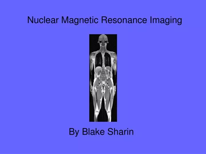
Nuclear Magnetic Resonance Imaging By Blake Sharin
Introduction • NMRI or MRI • Basics of MRI • Magnetic Intensity • Magnets • Nuclear Magnetic Resonance – the basic physics • Advantages and Disadvantages • Future of MRI
History • July 3, 1977 • First MRI exam produced on a human body • Dr. Raymond Damadian (physician and scientist) • 5 hours to produce one image • 7 years of labored work • Original machine called “Indomitable” • Spirit of their struggle to do what many people thought could not be done
Basics of MRI • MRI – Magnetic Resonance Imaging • imaging technique used primarily in medical settings to produce high quality images of the inside of the human body. • based on the principles of nuclear magnetic resonance (NMR). • a spectroscopic technique used by scientists to obtain microscopic chemical and physical information about molecules.
Basics of MRI (contd.) • NMRI • The technique was called magnetic resonance imaging rather than nuclear magnetic resonance imaging (NMRI) because of the negative connotations associated with the word nuclear in the late 1970's.
Magnetic Intensity • Biggest most important part • Magnet • Tesla • 0.5 – 2.0 Tesla • Over 2 have not been approved for medical use • 2 Tesla = 20,000 Gauss • Earth’s magnetic field is 0.5 Gauss
Magnetic Intensity (contd.) • Metal objects can become projectiles • paperclips, pens, keys, scissors, hemostats, stethoscopes and any other small objects can be pulled out of pockets and off the body without warning. • Credit Cards, bank cards and anything else with magnetic encoding will be erased by most MRI systems
Magnetic Intensity (contd.) • Magnetic Force • Force exerted on an object increases exponentially as it nears the magnet. • Example • Imagine standing 15 feet (4.6 m) away from the magnet with a large pipe wrench in your hand. You might feel a slight pull. Take a couple of steps closer and that pull is much stronger. When you get to within 3 feet (1 meter) of the magnet, the wrench likely is pulled from your grasp.
Magnets • There are 3 basic types of magnets used in MRI systems. • Resistive • Permanent • Superconducting
Resistive Magnet • Consists of many windings or coils of wire wrapped around a cylinder or bore through which an electric current is passed. • This causes a magnetic field to be generated. • If the electricity is turned off, the magnetic field dies out. • These magnets are lower in cost to construct than others. • Require huge amounts of electricity (up to 50 kilowatts) to operate because of the natural resistance in the wire. • To operate this type of magnet above about the 0.3-tesla level would be prohibitively expensive.
Permanent Magnet • Its magnetic field is always there and always on full strength, so it costs nothing to maintain the field. • The major drawback is that these magnets are extremely heavy. • They weigh many, many tons at the 0.4-tesla level. A stronger field would require a magnet so heavy it would be difficult to construct. • Permanent magnets are getting smaller, but are still limited to low field strengths.
Superconducting Magnets • By far the most commonly used. • Somewhat similar to a resistive magnet -- coils or windings of wire through which a current of electricity is passed create the magnetic field. • Important difference • wire is continually bathed in liquid helium at 452.4 degrees below zero. • Insulated • Superconductive systems are still very expensive, but they can easily generate 0.5-tesla to 2.0-tesla fields, allowing for much higher-quality imaging.
Protons produce a small magnetic field The Basic Physics A moving electric charge produces a magnetic field Protons have a positive charge Protons spin
Basic Physics (contd.) • The human body is made up of untold billions of atoms, the fundamental building blocks of all matter. • The nucleus of an atom spins on an axis. You can think of the nucleus of an atom as a top spinning somewhere off its vertical axis. • We are only concerned with the hydrogen atom. It is an ideal atom for MRI because its nucleus has a single proton
Proton Alignment I No external field… Randomly aligned
Proton Alignment II External field… Aligned with field
Advantages and Disadvantages • Diagnosing multiple sclerosis (MS) • Diagnosing tumors of the pituitary gland and brain • Diagnosing infections in the brain, spine or joints • Visualizing torn ligaments in the wrist, knee and ankle • Visualizing shoulder injuries • Diagnosing tendonitis • Evaluating masses in the soft tissues of the body • Evaluating bone tumors, cysts and bulging or herniated discs in the spine • Diagnosing strokes in their earliest stages
Advantages and Disadvantages (Contd.) • There are many people who cannot safely be scanned with MRI (for example, because they have pacemakers), and also people who are too big to be scanned. • There are many claustrophobic people in the world, and being in an MRI machine can be a very disconcerting experience for them. • MRI scans require patients to hold very still for extended periods of time. MRI exams can range in length from 20 minutes to 90 minutes or more.
Future of MRI • This technology is still in its infancy, comparatively speaking. • It has been in widespread use for less than 20 years (compared with over 100 years for X-rays). • Very small scanners for imaging specific body parts are being developed.
References • http://www.cis.rit.edu/htbooks/mri/ • http://www.medicinenet.com/MRI_Scan/article.htm