Download
1 / 40
420 likes | 625 Views
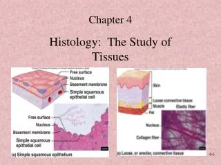
Preferential Utilization استعمال of Energy by Tissues. Dr. M. Azhar Chishti, Medical Biochemistry. Objective. To understand the different ways of energy utilization by various organs

E N D
Preferential Utilization استعمال of Energy by Tissues Dr. M. Azhar Chishti, Medical Biochemistry
Objective • To understand the different ways of energy utilization by various organs • To be familiar with: how fuel availability during absorptive state affects different organs’ energy utilization relative to fasting state.
Lecture Outline • 1. Overview of the major ways in which glucose is metabolized in selected tissues • 2. Inter-tissue relationships in absorptive and starvation states: • A- Liver • B- Brain • C- Skeletal muscle and heart • D- Adipose tissue • E- Red blood cells • F- Kidneys
Well-fed (absorptive) state vs Fasting/Starvation • During the absorptive الامتصاص state (which is the 2 to 4hour period after ingestion of a normal meal), virtually واقعيا , ALL tissues use glucose as a fuel. • While during fasting/starvation various organs use different fuels to obtain energy
continue • Metabolites المواد الايضيه move between tissues. • An “organ map” will be introduced in this lecture. • The goal of this “organ map” is to create an expanded and clinically useful vision of whole-body metabolism
Liver: “Nutrient الاغذيه distribution center” In well-fed (absorptive) state In fasting طريقه المحاضره بيعطيك اربع او خمس اعضاء كل عضو يعطيك اياه على فترتين ( الصيام والغذاء ) ويعلمك في كل فتره وش بيصير للكاربوهيدرات والبروتين والدهون
The liver is considered a “nutrient distribution center” • After a meal, the liver is bathed مغمور بـ in blood containing absorbed nutrients and elevated levels of insulin secreted by the pancreas. These reached the liver through the hepatic portal vein before entry into the general circulation. • Thus , the liver smoothes out potentially broad fluctuations تغير عريض in the availability of nutrients for the peripheral tissues.
Major metabolic pathways in liver in the absorptive state تلخص كل العمليات بالكبد # طريقه هذا الدكتور يعطيك صوره مثل هذي ثمن يشرح في الشرائح اللي بعدها
The liver is normally a glucose-producing rather than a glucose-using tissue. • However, after a meal containing carbohydrate, the liver becomes a net consumer of glucose. • What are the mechanisms responsible for this in hepatic metabolism of glucose after a meal containing carbohydrates? Next
Mechanisms responsible for increased hepatic metabolism of glucose after a meal containingcarbohydrates • Increased phosphorylation of glucose by glucokinase • Increased glycogen synthesis • Increased activity of hexose monophosphate pathway (HMP) • Increased glycolysis and decreased gluconeogenesis
Mechanisms affecting hepatic fat metabolism after a meal • Increased fatty acid synthesis • Increased triacylglycerol (TAG) synthesis packaged into very-low-density lipoprotein (VLDL) particles secreted into the blood to be used by extrahepatic tissues (particularly adipose & muscle tissue)
Mechanisms affecting hepatic amino acids metabolism after a meal • Increased amino acid degradation: • The excess amino acids; after the synthesis of proteins and other N2-containing molecules; are not stored, but are either: • Released into the blood for all tissues to use in protein synthesis (especially the branched-chain amino acids which cannot be degraded by the liver, and are preferentially metabolized in muscle) • Degraded to pyruvate, acetyl CoA, or TCA cycle intermediates energy production or fatty acid synthesis • Increased protein synthesis
Liver during fasting/starvation glycogen degradation gluconeogenesis KB synthesis FA oxidation
Carbohydrate metabolic response of the liver during fasting: • The primary role of liver during fasting is maintenance of blood glucose for use by other organs. • This takes place through the following mechanisms: • Increased glycogen degradation maintain blood glucose during early fasting, because liver glycogen store is nearly exhausted after 10-18 hrs of fasting. • Increased gluconeogenesis: starts 4-6 hrs after the last meal and becomes fully active as stores of liver glycogen are depleted; i.e. maintain blood glucose during prolonged fasting.
Fat metabolic response of the liver during fasting: • FA oxidation: FA are from adipose tissue and represent the major source of energy for the liver for postabsorptive state. • Ketone bodies synthesis: starts during the first days of fasting, can be used as fuel by most tissues, including the brain. Although KB synthesis occurs in the liver, the liver cannot use KB as fuels.
العضو الثاني Adipose Tissue: “Energy Storage Depot” In well-fed (absorptive) state In fasting
minimal source of FA in human adipose tissue Major sources of FA in human adipose tissue are:
Carbohydrate metabolism in adipose tissue in the absorptive state حاله الغذاء -الامتصاص • Increased glucose transport (insulin-dependent) • Increased glycolysis glycerol phosphate synthesis used in TAG synthesis • Increased activity of HMP NADPH
Fat metabolism in adipose tissue in the absorptive state • Increased synthesis of fatty acids • Increased TAG synthesis • Decreased TAG degradation (due to the insulin-mediated inhibition of hormone-sensitive lipase; HSL)
Carbohydrate metabolism in adipose tissue in Fasting Decreased glucose transport and metabolism (due to low levels of insulin) decreased FA & TAG synthesis
Fat metabolism in adipose tissue in Fasting • Increased TAG degradation (due to hormonal activation of HSL) • Increased FA release from TAG degradation. These FA are either: • released into blood to tissue to be used as fuel • converted to acetyl CoA in adipocytes TCA energy production • Decreased FA uptake (due to low activity of lipoprotein lipase in adipose tissue in fasting)
العضو الثالث Resting Skeletal Muscle In well-fed (absorptive) state In fasting
Energy metabolism of skeletal muscle Skeletal muscle is able to respond to substantial changes in the demand for ATP. At rest, muscle accounts for ~ 30% of the O2 consumption of the body. During vigorous muscle exercise, muscle accounts for up to 90% of the total O2 consumption
Carbohydrate metabolism in resting skeletal muscle in the absorptive state • Increased glucose transport (insulin-dependent) glucose metabolism energy for the muscle (Glucose is the 1ary source of energy for muscle in the well-fed state) • Increased glycogen synthesis
Amino acid metabolism in resting skeletal muscle in the absorptive state • Increased protein synthesis (to replace proteins degraded since the previous meal) • Increased uptake of branched-chain amino acids used for: • protein synthesis & • source of energy
Fat metabolism in resting skeletal muscle in the absorptive state • FA are of secondary importance as a fuel for muscle during the absorptive state. • In the absorptive (well-fed) state; glucose is the primary source of energy for skeletal muscle.
Carbohydrate metabolism in resting skeletal muscle in Fasting Depressed glucose uptake and metabolism in skeletal muscle in fasting (due to low levels of circulating insulin)
Amino acid metabolism in resting skeletal muscle in Fasting • During the first few days of fasting rapid protein breakdown (muscle proteolysis) AA to be used by the liver for gluconeogenesis maintain adequate blood glucose level for brain energy requirements • Several weeks of fasting decreased rate of muscle proteolysis (the brain has begun using KB as a source of energy
Fatmetabolism in resting skeletal muscle in Fasting • First 2 weeks of fasting: the source of energy for muscle are: • FA oxidation (FA are from adipose tissue) • KB from liver • After ~ 3 weeks of fasting: the source of energy for muscle is: • FA oxidation (FA are from adipose tissue) (while KB are spared for the use of brain as a fuel)
Differences between heart muscle and skeletal muscle energy requirements • Heart muscle differs from skeletal muscle in three important ways: • the heart is continuously active, whereas skeletal muscle contracts intermittently on demand; • the heart has a completely aerobic metabolism • the heart contains negligible تافه - قليل energy stores, such as glycogen or lipid. • Thus, any interruption of the vascular supply, for example, as occurs during a myocardial infarction, results in rapid death of the myocardial cells. • Fuels used by heart muscle • Glucose • free fatty acids (FFA) • ketone bodies (KB)
االعضو الرابع Brain In well-fed (absorptive) state In fasting
Carbohydrate & fat metabolism in brain in the absorptive state • The brain completely depends on the availability of blood glucose as a fuel. • Little contribution of FA to energy production to brain (FA do not efficiently cross the blood-brain barrier) ~140g glucose/day
Carbohydrate & fat metabolism in brain in the Fasting/starvation state • During the first few days of fasting: The brain continues to use blood glucose exclusively as a fuel. • Prolonged fasting (> 2-3 weeks) plasma Ketone bodies replace glucose as the 1ary fuel for the brain.
العضو الخامس Red Blood Cells
Red Blood Cells (RBC) • Glucose is the only fuel source for RBC • Glucose enters RBS through GLUT-1 (insulin-independent) • Glucose is then metabolized as follows: • mainly by glycolysis to produce energy & 2,3 Bisphosphoglycerate (Anaerobic glycolysis since RBC has no mitochondria) lactate into blood • HMP NADPH Protective antioxidant function
ملخص لجميع الاعضاء وربط العلاقه فيما بينهما حاله الغذاء
هذا مسجد مهم جدااا ( سؤااال ) Thank you Thank You