Download
1 / 10
100 likes | 214 Views
Curvature effect on the Structure of Phospholipid Bilayers Norbert Ku č erka NSERC V isiting Fellow in CNBC – NRC. Collaborators. Jeremy Pencer John Katsaras Mu-Ping Nieh CNBC – NRC, Chalk River, ON John Nagle Carnegie Mellon University, Pittsburgh, PA Jonathan Sachs

E N D
Curvature effect on theStructure of Phospholipid BilayersNorbert KučerkaNSERC Visiting Fellow in CNBC – NRC
Collaborators • Jeremy Pencer • John Katsaras • Mu-Ping Nieh • CNBC – NRC, Chalk River, ON • John Nagle • Carnegie Mellon University, Pittsburgh, PA • Jonathan Sachs • University of Minnesota, Minneapolis, MN
Outline • Motivation • Experimental design • Effect of Vesicle Size • Bilayer Asymmetry • Conclusions • Outlook
Motivation A combined global analysis approach takes advantage of the complementarity of the two sample preparations, thus enhancing the spatial resolution of the bilayer structure. However, the method of combining two different data sets raises an important question: Do the flat lamellae and curved vesicles comprise equivalentbilayers? N. Kučerka, Y. Liu, N. Chu, H. I. Petrache, S. Tristram-Nagle, and J.F. Nagle, Biophys. J (2005)
Experimental design Bilayer curvature was varied as a function of ULV diameter, resulting from the extrusion through filter sizes of 500, 1000 and 2000 Å. Systems of pure DOPC ULVs are generally contaminated with pauci-lamellar vesicles. Extruded ULVs were characterized by DLS to determine their size distributions. Values for the two nominally smaller ULVs were shifted toward larger sizes (620 and 1210 Å), while it was reverse (1840 Å) for the largest one. The production of large ULVs was achieved by the addition of small amounts (up to 4 mol %) of the charged lipid, DOPS.
(NO) Effectof Vesicle Size The direct comparison of experimental data. SANS The SANS data revealed no differences in the mid-q scattering range that corresponds to the overall thickness of the bilayer. SAXS The most important “fingerprints” of the SAXS data consist of the positions of the scattering curve minima and maxima, that provide intrabilayer information. Good agreement of the different curves implies again no difference in bilayer structure.
(NO) Effectof Curvature SAXS form factors for 620-Å-diameter ULVs (red squares) compared to oriented multibilayers (blue diamonds), both made up of neutral DOPC lipid. Agreement between the two data sets suggests no difference in the structure of flat (Ø~) or curved (Ø~600 Å) bilayer. N. Kučerka, J. Pencer, J. N. Sachs, J. F. Nagle and J. Katsaras, Langmuir (2007)
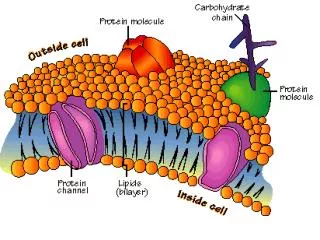
Bilayer Asymmetry The real space structure (scattering density profile) of a bilayer and reciprocal space scattering data are related through the Fourier transform. For a symmetric bilayer it is given as which is responsible for typical extinctions in the scattering intensity, as was observed in a case of DOPC bilayer. On the other hand, a lack of zero intensity indicates bilayer asymmetry, as can be deduced from the complete form of the Fourier transform This is exactly what we observed in the case of chargedDOPS bilayers.
Symmetry vs. Asymmetry There is no asymmetry found in DOPC bilayers, as is indicated by the scattering curves that approach zeros in the vicinity of scattering minima. On the other hand, charged DOPS lipids result in the formation of an asymmetric bilayer. This is readily detectable because of the distinct scattering effects associated with this bilayer morphology. N. Kučerka, J. Pencer, J. N. Sachs, J. F. Nagle and J. Katsaras, Langmuir (2007)
Conclusions • Both SANS and SAXS data showed no curvature effect on the bilayer structure ofneutral, zwitterionic lipids (DOPC). • Electrostatic interactions present in ULVs composedof charged DOPS lipids result in the formation of an asymmetric bilayer. • Theelectrostatic effects affecting the structure of charged bilayer and those due to the curvature are coupled. • The curvature alone does not induce any changes to the bilayer structure for ULVs larger than 600 Å.