Download
1 / 16
1.27k likes | 5.46k Views
Bronchiectasis. Dr.Samet.M Yazd University Harrison's PRINCIPLES OF INTERNAL MEDICINE-7th Edition. Definition. Bronchiectasis is an abnormal and permanent dilatation of bronchi It may be either focal or diffuse It is a disorder that typically affects older individuals

E N D
Bronchiectasis Dr.Samet.M Yazd University Harrison'sPRINCIPLES OF INTERNAL MEDICINE-7th Edition
Definition • Bronchiectasis is an abnormal and permanent dilatation of bronchi • It may be either focal or diffuse • It is a disorder that typically affects older individuals • approximately 2/3 of patients are women
Pathology • bronchial dilatation is associated with destructive and inflammatory changes in the walls of medium-sized airways, often at the level of segmental or subsegmental bronchi • Airway inflammation is primarily mediated by neutrophils and results in up-regulation of enzymes such as elastase and matrix metalloproteinases • The normal structural components of the wall, including cartilage, muscle, and elastic tissue, are destroyed and may be replaced by fibrous tissue • The dilated airways frequently contain pools of thick, purulent material, while more peripheral airways are often occluded by secretions or obliterated and replaced by fibrous tissue • Additional microscopic features include bronchial and peribronchial inflammation and fibrosis, ulceration of the bronchial wall, squamous metaplasia, and mucous gland hyperplasia • The parenchyma normally supplied by the affected airways is abnormal, containing varying combinations of fibrosis, emphysema, bronchopneumonia, and atelectasis
Pathology • As a result of inflammation, vascularity of the bronchial wall increases, with associated enlargement of the bronchial arteries and anastomoses between bronchial & pulmonary arterial circulations
patterns of bronchiectasis Three different patterns of bronchiectasis have been described • cylindrical bronchiectasis: the involved bronchi appear uniformly dilated and end abruptly at the point that smaller airways are obstructed by secretions • varicose bronchiectasis: the affected bronchi have an irregular or beaded pattern of dilatation resembling varicose veins • saccular (cystic) bronchiectasis: the bronchi have a ballooned appearance at the periphery, ending in blind sacs without recognizable bronchial structures distal to the sacs
Etiology and Pathogenesis Infection is the usual cause of the inflammation • microorganisms such as Pseudomonas aeruginosaand Haemophilus influenzaeproduce pigments, proteases, and other toxins that injure the respiratory epithelium and impair mucociliary clearance the dilated airways become more susceptible to colonization and growth of bacteria a reinforcing cycle can result, with inflammation producing airway damage, impaired clearance of microorganisms, and further infection, which then completes the cycle by inciting more inflammation.
Infectious Causes • Adenovirus and Influenza virus are the main viruses that cause bronchiectasis in association with lower respiratory tract involvement • Virulent bacterial infections, especially with potentially necrotizing organisms such as Staphylococcus aureus, Klebsiella, and Anaerobes • Infection with Bordetellapertussis, particularly in childhood • Bronchiectasis has been reported in patients with HIV infection • Tuberculosis • Nontuberculousmycobacteriacan serve as primary pathogens & secondary infections or colonizing organisms Impaired host defense mechanisms are often involved in the predisposition to recurrent infections • The major cause of localized impairment of host defenses is endobronchial obstruction • Slowly growing endobronchialneoplasms such as carcinoid tumors may be associated with bronchiectasis • Foreign-body aspiration is another important cause of endobronchial obstruction, particularly in children • Airway obstruction can also result from bronchostenosis, from impacted secretions or extrinsic compression by enlarged lymph nodes • Generalized impairment of pulmonary defense mechanisms occurs with: • immunoglobulin deficiency • primary ciliary disorders • cystic fibrosis (CF) • With panhypogammaglobulinemia patients often also have a history of sinus or skin infections • Selective deficiency of an IgG subclass, especially IgG2, has also been described in a small number of patients with bronchiectasis.
primary ciliarydyskinesia • are responsible for 5–10% of cases of bronchiectasis • is inherited in an autosomal recessive fashion • Numerous defects are encompassed under this category, including structural abnormalities of the dynein arms, radial spokes, and microtubules; mutations in heavy and intermediate chain dynein have been described in a small number of patients • The cilia become dyskinetic; their coordinated, propulsive action is diminished, and bacterial clearance is impaired • clinical effects include recurrent upper and lower respiratory tract infections, such as sinusitis, otitis media, and bronchiectasis • males are generally infertile visceral rotation during development depends upon proper ciliary motion, positioning of normally lateralized organs becomes random approximately half of patients fall into the subgroup of Kartagener's syndrome • situsinversus • bronchiectasis • sinusitis
Noninfectious Causes Some cases of bronchiectasis are associated with exposure to a toxic substance • inhalation of a toxic gas such as ammonia or aspiration of acidic gastric contents • An immune response in the airway may also trigger inflammation, destructive changes, and bronchial dilatation ABPA • α1-antitrypsin deficiency »»»»»»»»»»»»»»»»panacinar emphysema & bronchiectasis • Yellow nail syndrome which is due to hypoplastic lymphatics, is accompanied by bronchiectasis in approximately 40% of patients: • lymphedema • pleural effusion • yellow discoloration of the nails
Clinical Manifestations • Patients typically present with persistent or recurrent cough and purulent sputum production • Hemoptysis occurs in 50–70% of cases • More significant, even massive bleeding is often a consequence of bleeding from hypertrophied bronchial arteries • Systemic symptoms such as fatigue, weight loss, and myalgias can also occur • When a specific infectious episode initiates bronchiectasis, patients may describe a severe pneumonia followed by chronic cough and sputum production • patients without a dramatic initiating event often describe the insidious onset of symptoms • some cases are either asymptomatic or have a nonproductive cough »»»»»»»»»»» dry bronchiectasis in an upper lobe • Dyspnea or wheezing generally reflects either widespread bronchiectasis or underlying COPD • With exacerbations of infection, the amount of sputum increases, becomes more purulent and often more bloody; systemic symptoms, including fever, may also be prominent. Physical examination : • Any combination of crackles, rhonchi, and wheezes may be heard • clubbing may be present • cor pulmonale and right ventricular failure
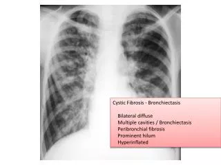
Radiographic • the findings of chest radiograph are often nonspecific • radiograph may be normal with mild disease • saccular bronchiectasis may have prominent cystic spaces,with or without air-liquid levels, corresponding to the dilated airways dilated airways + thickened walls • When seen longitudinally, the airways appear as "tram tracks“ • when seen in cross-section, they produce "ring shadows" • Because the dilated airways may be filled with secretions, the lumen may appear dense rather than radiolucent, producing an opaque tubular or branched tubular structure. • HRCT images 1.0–1.5 mm thick, provides an excellent view of dilated airways it is now the standard technique for detecting or confirming the diagnosis of bronchiectasis
Laboratory Findings • Examination of sputum • When bronchiectasis is focal, FOB may reveal an underlying endobronchial obstruction • upper lobe involvement may be suggestive of either TB or ABPA • With more widespread disease, measurement of sweat chloride levels for CF, structural or functional assessment of nasal or bronchial cilia or sperm for primary ciliary dyskinesia, and quantitative assessment of immunoglobulins may explain recurrent airway infection • PFT tests may demonstrate airflow obstruction • Bronchial hyperreactivityto methacholine challenge, and some reversibility of the airflow obstruction with inhaled bronchodilators are relatively common the workup should be dictated by a careful assessment of the clinical scenario »»»»»»»»» • patient with focal bronchiectasis documentation of a prior pneumonia in same location may suffice • Evaluation for immunoglobulin deficiency and CF should be considered for young patients with bronchiectasis and sino-pulmonary disease
Treatment • Therapy has several major goals: (1) treatment of infection, particularly during acute exacerbations (2) improved clearance of tracheobronchial secretions (3) reduction of inflammation (4) treatment of an identifiable underlying problem Antibiotics are the cornerstone of bronchiectasis management • antibiotics are used only during acute episodes • choice of an antibiotic should be guided by Gram's stain and culture of sputum • empiric coverage (amoxicillin, co-trimoxazole,levofloxacin) is often given initially • Infection with P. aeruginosais of particular concern, as it appears to be associated with greater rate of deterioration of lung function and worse quality of life • There are no firm guidelines for length of therapy, but a 10–14 day course or longer is typically administered facilitate drainage : mechanical methods and devices & appropriate positioning • Mucolytic agents to thin secretions and allow better clearance are controversial • Aerosolized recombinant DNase, which decreases viscosity of sputum by breaking down DNA released from neutrophils, has been shown to improve pulmonary function in CF but may be deleterious and should be avoided in bronchiectasis not associated with CF • Bronchodilators to improve obstruction and aid clearance of secretions are useful in patients with airway hyperreactivity and reversible airflow obstruction surgical therapy »»»»»»»»»»»»»»»»»»» when bronchiectasis is localized and the morbidity is substantial despite adequate medical therapy massive hemoptysis, often originating from the hypertrophied bronchial circulation • conservative therapy, including rest and antibiotics • surgical resection • bronchial arterial embolization • Although resection may be successful if disease is localized, embolization is preferable with widespread disease