Download
1 / 20
240 likes | 770 Views
PATIENT POSITIONING IN NEUROANAESTHESIA. Dr. Rahul Norawat. University College of Medical Sciences & GTB Hospital, Delhi. INTRODUCTION. Positioning is the joint responsibility of the surgeon & anesthesiologist.

E N D
PATIENT POSITIONING IN NEUROANAESTHESIA. Dr. RahulNorawat University College of Medical Sciences & GTB Hospital, Delhi
INTRODUCTION • Positioning is the joint responsibility of the surgeon & anesthesiologist. • Ideal pt. positioning involves balancing surgical comfort, against the risks related to the pt. position. • Pt. positioning & postural limitation should be considered during the PAC.
Positioning of neurosurgical patient: • Adequate anesthetic depth, • Hemodynamic stability, • Oxygenation, • Preservation of monitors. • Disconnection - create “blackout” state. • ASA task force general guidelines.
Head Positioning Ideal position of head for Craniotomies & spine procedures based on the 2 principles: • An imaginary trajectory from the highest point at skull surface to area of interest in brain should be the shortest distance between the 2 points. • The exposed surface of the skull & an imaginary perimeter of craniotomy should be parallel to the floor.
Types of Craniotomies • Ant. Parasagittal • Frontosphenotemporal • Sub-temporal • Lat Sub-occipital • Midline Sub-occipital • Post. Parasagittal
Fixation of the Head For craniotomies or burr holes, head positioned on: • Horseshoe headrest (doughnut), • Skeletally fixed with 3 (Mayfield frame) or 4-pins fixation device.
Local infiltration • iv anesthetic agent (propofol 0.5-1 mg/kg) • Inhalational anesthetic Application of a skeletal fixation pins Tachycardia and hypertension Rupture of untreated cerebral aneurysms • Benefits : Immobility, surgical comfort. • Risks : Bleeding, air embolism, scalp and eye laceration, pressure alopecia.
Head and Neck positioning: Rotation, Hyperflexion, Hyperextension Brain stem & cervical spine ischemia. Quadriparesis, quadriplegia & cerebral infarction.
At risk Patients : • Osteophytes, • Arthritis, • Vascular atherosclerosis. • Head safely rotated b/w 0-45°. • For more rotation, a roll/pillow place under the opposite shoulder. • Maintaining 2-3 finger breadths thyromental distance during neck flexion.
Benefits: Surgical comfort & optimal access to the surgical area. • Risks & complications: • Postoperative discomfort & pain, • Brachial plexus injury, • Obstruction of : • Jugular veins & vertebral venous plexuses, • Cerebral lymphatic, • Vertebral or carotid arteries. • CSF flow,
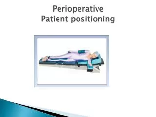
Body Positioning • Supine Position (Dorsal Decubitus) • Most frequently utilized position. • Used for : • Cranial procedures, • Carotid endarterectomies, • Ant. approaches to cervical & lumbar spine.
A Horizontal position : poorly tolerated. B Lawn chair position : 15° angulation's & flexion at trunk-thigh-knee & more physiological positioning of lumbar spine, hips and knees. C Reverse Trendelenbourg position : 10-15° repositioning from the horizontal axis. A - horizontal position, B - lawn chair (contoured) position, C - reverse Trendelenbourg position.
Arms: • Abducted: < 90° to minimize brachial plexus injury. • Adducted • Hand & forearm: • Supinated or neutral position to reduce external pressure on spiral groove of humerus & ulnar nerve.
Benefits: • Simplest, • Not require special instrumentation & • Not require disconnection of tracheal tube & invasive monitors. • Risks: • Head rotation or flexion for optimal surgical conditions, • Pressure alopecia, • Pressure point reaction, • Nerve injury. cubital tunnel retinaculum (CTR)
Hemodynamic and Ventilation. • Every 2.5 cm change of vertical ht. from the reference point at level of the heart leads to a change of MAP by 2 mmHg in the opposite direction. • V & Q are best in dependent lungs. • Positive-pressure ventilation provides the best ventilation to non-dependent lung zones -V/Q mismatch.
B. Lateral Position • Temporal lobe craniotomy, • Skull base, • Posterior fossa procedures & • Retroperitoneal approach to thoracolumbar spine.
B A A - Dependent arm is hung under the operating table, an upper arm is placed on the arm board, B - Dependent arm is positioned on the operating table and an arm board, an upper arm is placed over the trunk on the pillow.
Park-bench position: • Modification of lat. position. • Better access to posterior fossa. • Upper arm positioned along lateral trunk & upper shoulder is taped towards table. Hemodynamic and Ventilation. • In awake patient, Zone 3 West is occupying the dependent 18 cm of lung tissue. Lung tissue above 18 cm from bed level is not perfused. • During GA & positive pressure ventilation, the non-dependent lung zones are ventilated better - worsening V/Q mismatch.