Download

1 / 96
1k likes | 1.26k Views
Hemorrhage & Shock. Terry White, RN. Review of Hemorrhage. Location Anatomical Type & Timing Coagulation Fibrinolysis Assessment Management. Review of Hemorrhage. Location External Internal Traumatic Non-Traumatic Examples?. Review of Hemorrhage. Anatomical Type Arterial Venous

E N D
Hemorrhage & Shock Terry White, RN
Review of Hemorrhage • Location • Anatomical Type & Timing • Coagulation • Fibrinolysis • Assessment • Management
Review of Hemorrhage • Location • External • Internal • Traumatic • Non-Traumatic • Examples?
Review of Hemorrhage • Anatomical Type • Arterial • Venous • Capillary • Timing • Acute • Chronic
Severity of Hemorrhage Comparison of Adult vs Child
Hematocrit • % of RBC in blood (hematocrit) • Normal: • 37% - 47% (Female) • 40% - 54% (Male)
Thrombocytes • Platelets • Form platelet plugs • contact collagen & adhere to injured surface • activate platelets • aggregate to form platelet plug
Coagulation • Formation of blood clots • Prothrombin activator • Prothrombin Thrombin • Fibrinogen Fibrin • entrap platelets, blood cells & plasma • Clot retraction
Fibrinolysis • Breaking up the clot • tissue plasminogen activator (tPA) • plasminogen plasmin
Assessing Hemorrhage • Clues • Bright red blood from wound, mouth, rectum or other orifice • Hematemesis • Coffee ground appearance of vomitus • Hematochezia • Melena • Orthostatic hypotension • Dizziness or syncope on sitting or standing • Signs and symptoms of hypovolemic shock
Management of Hemorrhage • Airway and Ventilatory Support • Circulatory Support • From nose or ears after head trauma = loose drsg • Control bleeding • direct pressure, elevation, pressure points • tourniquet • packing of large wounds • splinting • PASG • transport to appropriate facility
Shock “A rude unhinging of the machinery of life” “A brief pause in the act of dying”
Shock Inadequate peripheral perfusion leading to failure of tissue oxygenation may lead to anaerobic metabolism
Shock • Homeostasis • cellular state of balance • perfusion of cells with oxygen is one of its cornerstones
Shock • Adequate Cellular Oxygenation • Red Cell Oxygenation • Red Cell Delivery To Tissues Fick Principle
The following variables are measured: • VO2 consumption per minute using a spirometer (with the • subject re-breathing air) and a CO2 absorber • Cv, the oxygen content of blood taken from the • pulmonary artery (representing deoxygenated blood • blood) • Ca, the oxygen content of blood from a cannula in a • peripheral artery (representing oxygenated blood) From these values, we know that: where CO = Cardiac Output, CA = Oxygen concentration of arterial blood and CV = Oxygen concentration of venous blood. ( x CV ) x CA ) CO = ( CO VO2 This allows us to say VO2 CO = CA CV
Fick Principle Air’s gotta go in and out. Blood’s gotta go round and round. Any variation of the above is not a good thing!
Shock • Red Cell Oxygenation • Oxygen delivery to alveoli • Adequate FiO2 • Patent airways • Adequate ventilation
Shock • Red Cell Oxygenation • Oxygen exchange with blood • Adequate oxygen diffusion into blood • Adequate RBC flow past alveoli • Adequate RBC mass/Hgb levels • Adequate RBC capacity to bind O2 • pH • Temperature
Shock • Red Cell Delivery To Tissues • Adequate perfusion • Blood volume • Cardiac output • Heart rate • Stroke volume (pre-load, contractility, after-load) • Conductance • Arterial resistance • Venous capacitance
Shock • Red Cell Delivery To Tissues • Adequate RBC mass • Adequate Hgb levels • Adequate RBC capacity to unbind O2 • pH • Temperature • Distance between capillaries and cells
Shock Inadequate oxygenation or perfusion causes: Inadequate cellular oxygenation Shift from aerobic to anaerobic metabolism
6 CO2 6 O2 6 H2O METABOLISM 36 ATP GLUCOSE HEAT (417 kcal) AEROBIC METABOLISM Glycolysis: Inefficient source of energy production; 2 ATP for every glucose; produces pyruvic acid Oxidative phosphorylation: Each pyruvic acid is converted into 34 ATP
2 LACTIC ACID GLUCOSE METABOLISM 2 ATP HEAT (32 kcal) ANAEROBIC METABOLISM Glycolysis: Inefficient source of energy production; 2 ATP for every glucose; produces pyruvic acid
Anaerobic Metabolism • Occurs without oxygen • oxydative phosphorylation can’t occur without oxygen • glycolysis can occur without oxygen • cellular death leads to tissue and organ death • can occur even after return of perfusion • organ or organism death
Inadequate Cellular Oxygen Delivery Lactic Acid Production Inadequate Anaerobic Energy Production Metabolism Metabolic Failure Metabolic Acidosis CELL DEATH Ultimate Effects of Anaerobic Metabolism
Maintaining perfusion requires: • Volume • Pump • Vessels • Failure of one or more of these causes shock
Shock • Hypovolemic Shock = Low Volume • Trauma • Non-traumatic blood loss • Vaginal • GI • GU • Burns • Diarrhea • Vomiting • Diuresis • Sweating • Third space losses • Pancreatitis • Peritonitis • Bowel obstruction
Shock • Cardiogenic Shock = Pump Failure • Mechanical obstruction (“distributive shock”) • Cardiac tamponade • Tension pneumothorax • Pulmonary embolism • Acute M I • CHF • Bradyarrhythmias • Tachyarrhythmias
Shock • Vasogenic Shock = Low Resistance • Spinal cord trauma • neurogenic shock • Depressant drug toxicity • Simple fainting
Shock • Mixed Shock • Septic Shock • Overwhelming infection • Inflammatory response occurs • Blood vessels • Dilate (loss of resistance) • Leak (loss of volume)
Shock • Mixed Shock • Septic Shock • Fever • Increased O2 demand • Increased anaerobic metabolism • Bacterial toxins • Impaired tissue metabolism
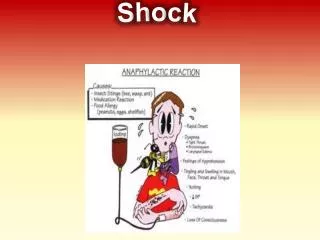
Shock • Mixed Shock • Anaphylactic Shock • Severe allergic reaction • Histamine is released • Blood vessels • Dilate (loss of resistance) • Leak (loss of volume)
Shock • Mixed Shock • Anaphylactic Shock • Histamine release • Extravascular smooth muscle spasm • Laryngospasm • Bronchospasm
Shock • Progressive syndrome • Three phases • Compensated • Decompensated • Irreversible
Shock • Signs and symptoms due to: • Hypoperfusion • Compensatory responses
Compensated Shock • Baroreceptors detect fall in BP • Usually 60-80 mm Hg (adult) • Sympathetic nervous system activates • What are the primary SNS Neurotransmitters & their effects?
Compensated Shock • Cardiac effects • Increased force of contractions • Increased rate • Increased cardiac output
Compensated Shock • Peripheral effects • Arteriolar constriction • Pre-/post-capillary sphincter contraction • Increased peripheral resistance • Shunting of blood to core organs
Compensated Shock • Decreased renal blood flow • Renin released from kidney arteriole • Renin & Angiotensinogen combine • Converts to Angiotensin I • Angiotensin I converts to Angiotensin II • Peripheral vasoconstriction • Increased aldosterone release (adrenal cortex) • promotes reabsorption of sodium & water
Compensated Shock • Decreased blood flow to hypothalamus • Release of antidiuretic hormone (ADH or Arginine Vasopressin) from posterior pituitary • Retention of salt, water • Peripheral vasoconstriction
Compensated Shock • Insulin • secretion caused by epinephrine • contributes to hyperglycemia • Glucagon • release caused by epinephrine • promotes liver glycogenolysis & gluconeogenesis • ACTH • stimulates adrenal cortex release of cortisol • glucose production
Compensated Shock • Peripheral capillaries contain minimal blood • Stagnation • Aerobic metabolism changes to anaerobic • Extracellular potassium shifts begin
Compensated Shock • Presentation • Restlessness, anxiety • Earliest sign of shock • Tachycardia • ?Bradycardia in cardiogenic, neurogenic
Compensated Shock • Presentation • Normal BP, narrow pulse pressure • Falling BP = late sign of shock • Mild orthostatic hypotension (15 to 25 mm Hg) • “Possible” delay in capillary refill
Compensated Shock • Presentation • Pale, cool skin • Cardiogenic • Hypovolemic • Flushed skin • Anaphylactic • Septic • Neurogenic
Compensated Shock • Presentation • Slight tachypnea • Respiratory compensation for metabolic acidosis
Compensated Shock • Presentation • Nausea, vomiting • Thirst • Decreased body temperature • Feels cold • Weakness
Decreased Cardiac Output Catecholamine Release Aldosterone, ADH Release Increased Blood Volume Increased PVR Increased Cardiac Output Increased Myocardial Work, O2 Demand Increased Volume Loss Compensated Shock Leading to Decompensation Myocardial Ischemia
Decompensated Shock • Presentation • Cardiac Effects • Decreased RBC oxygenation • Decreased coronary blood flow • Myocardial ischemia • Decreased force of contraction