Download
1 / 35
360 likes | 521 Views
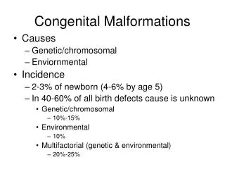
Congenital deformations . Developmental dysplasia of the hip. Congenital muscular torticolis. Clubfoot. Congenital muscular torticolis.

E N D
Congenital deformations.Developmental dysplasia of the hip. Congenital muscular torticolis. Clubfoot.
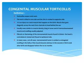
Congenital torticollis is a deformity of the neck characterised by tilting of the head to one side. There is a rotation of the occiput to one side and the chin to the opposite side.
The basic feature of this condition is a contracture of the sternomastoid muscle on one side. The exact pathology leading to the contracture is not known. It may be a myodysplasia of genetic origin.
Clinical features • The child may be brought soon after birth when the mother notices the tilt in the head to one side and often a swelling in the neck. There may be a firm swelling in the junction of the middle and distal third of the stemo-mastoid muscle. This is called the sternomastoid tumour. In cases due to birth trauma, it may be associated with fractures of the clavicle or Erb's palsy. In a left sternomastoid torticollis, the chin is tilted upwards to the right side and the occiput is tilted down on the left side.
Radiography • Radiography may show congenital anomalies in the cervical spine like hemivertebrae or partial fusion of the cervical vertebrae in cases where the stemomastoid is normal. In neglected cases, the contracture is severe and cannot be stretched passively. There is facial asymmetry, as the face in the affected side is not well developed. A new macula develops in the fundus to make the vision level in the tilted position of the head.
Radiography • In older children torticolis is due to the rotatory subluxation which may be due to infection, trauma
Treatment • A position in bed. • In the newborn stage, the stemomastoid is stretched passively by the physiotherapist. • After 4-6 weeks the child is taught neck exercises. • The treatment in later cases is mainly surgical tenotomy of the lower end of the stemomastoid.
There are 2 types of congenital dislocation of the hip. In the more common type, excessive laxity of the hip joint capsule and ligaments result in subluxation and dislocation. • In the other type, dysplasia or poor development of the acetabulum is the cause for the dislocation.
Clinical Features Ina new born child suspected to have congenital dislocation of the hip, look for any obvious shortening of the leg or any additional crease in the posterior dislocation. Two tests are performed. With the baby on its back, the hip and knee are held flexed and the limb is slowly abducted at the hip. If the hip is dislocated, the head of the femur is felt to slip in to the acetabulum with a click. This is the Ortolani's test. Barlow's test is a slightly modified one in which the head could be pushed in and out of the acetabulum with a click.
Clinical Features With the child lying on its back, the hip and knee are held flexed to 90 degrees, the pelvis is steadied with the left hand and the knee is held in the right hand. On pushing down and pulling up at the knee vertically, the head could be felt by the left hand to move down and up in the gluteal region. This is the telescopy test.
Clinical Features The condition often escapes notice till the child begins to walk when a limp is obvious. On examination, there is limitation of abduction of the flexed hip. The dislocated head of the femur can be palpated in the gluteal region. When the hips are flexed to 90 degrees and the feet rested on the table, the level of the knee of the affected leg is lower. This is called the Galeazzi's sign. The gait will be unstable. This is called the Trendelenberg gait due to the dislocated hip telescoping upward on putting weight on the leg.
Bilateral dislocation • There will be no difference between the two legs in length. The perineum will be wider than normal. Telescoping will be present in both hips. The older child walks with a gait swaying from side to side with a marked increase in the lordosis of the lumbar spine (Sailor's gait).
Radiological features • In the newborn baby, before the femoral epiphysis has appeared, the condition is to be suspected if the joint space is excessively broad and the neck of the femur lies far away from the acetabulum. In A.P. view of pelvis with both hips in abduction, normally a line drawn along the shaft of the femur prolonged proximally will pass through the centre of the acetabulum. When the hip is dislocated, this line will pass above the acetabulum. More recently ultra sound scanning is used to detect the position of the unossified cartilaginous head. In a child aged one year and above, the dislocated side shows the epiphysis of the head lying outside the acetabulum and at a higher level. The roof of the acetabulum slopes upwards when compared to the normal side.
Treatment • The treatment should be started as soon as possible after birth. The aim of the treatment is to reduce the hip and maintain it in the reduced position till it remains stable. The treatment is age related and depends on the age at which the child is brought. • In Infants below 3 months Splints like Von Rosen's splints are applied to retain the position of the head.
Treatment Children from 3 to 6 months • The dislocation is reduced by gentle manipulation under general anaesthesia and immobilised in plaster cast, with the hip at 90 degree flexion and 45 degree abduction. This is the "human position" as described by Kumar. The hips is left in an older should not be immobilised in the extreme position of the so called Frog position of 90 degrees • flexion and 90 degrees abduction. The excessive stretching of the capsule in this position interferes with the blood supply to the capital epiphysis.
Treatment Children from 6 to 12 months • A preliminary traction and gradual abduction is done for 2-3 weeks before reduction under general anaesthesia. Some of these children may need adductor tenotomy if there is resistance to reduction by its tightness. The plaster cast is then applied. • Children 1 to 3 years • In this age group, after preliminary traction. surgery will be required to reduce the head of femur into the acetabulum. In cases with excessive valgus and anteversion of the femoral neck, a femoral varus derotation osteotomy is done as a second stage. • Children 3 to 6 years • In this age group, after open reduction of the hip and femoral osteotomy, an osteotomy of the acetabulum is made to retain the head in stable position.
Neglected congenital dislocation of the hip • Neglected congenital dislocation of the hip may present after the age of 10 years with pain in the hip and unstable gait. These will require pelvic stabilising operations like Schantz osteotomy. • Complications • Avascular necrosis of the head of the femur is a complication of forceful reduction of the hip in childhood. Neglected cases develop painful instability of the hip and in later years osteoarthrosis of the hip.
Club Foot (CONGENITAL TALIPES EQUINO VARUS)
Congenital talipes equino varus is one of the most common congenital deformities in India when compared to the Western countries. The word Talipes is derived from 'Talus' and 'Pes' and was applied to those walking on their neglected deformities wherein the talus rested on the ground as the foot (Pes). It is characterised by a foot plantar flexed at the ankle, inverted at the subtalar joint and adducted at the forefoot. The deformity is bilateral in approximately 50% of cases and is more common in males.
Etiology • The exact cause of this deformity is not known. According to Hippocrates, the talipes deformity is of mechanical origin and is caused by an abnormal intra uterine position of the fetus. Another theory is that a primary germ plasm defect in the talus causes continued plantar flexion and inversion of this bone with subsequent soft tissue changes in the joints and musculo tendinous corn-plexus. The third hypothesis is that primary soft tissue abnormalities within the neuromuscular units cause secondary bony changes.
Types of CTEV • One type is a less severe deformity with an elevated heel and is mobileand is comparatively easy to correct by manipulation. This is called the extrinsic type. • The other type is a severe deformity with a small heel and extremely resistant to treatment and tends to relapse. This is the intrinsic type (so called vicious feet).
Pathological Anatomy • The structural changes are most evident in the talus and the calcaneum. The talus is plantar flexed and the superior articular surface is exposed. The neck becomes elongated and deflected medially and downwards. The calcaneum is inverted so that its medial tuberosity approaches the medial malleolus and its posterior aspect is elevated. The navicular is rotated on its axis and its tubercle may almost come into contact with the medial malleolus. • The soft tissues on the posterior aspect of the ankle and medial aspect of the foot are contracted. The tendo achilles, the tibialis posterior, the plantar fascia, the spring ligament, the deep plantar ligaments, flexor hallucis longus, flexor digitorum longus and abductor hallucis are contracted. The capsules of the subtaloid, talo navicular, naviculo cunieform joints are also contracted.
Clinical features The shape and contour of the foot is characteristic. There is equinus deformity at the ankle, varus deformity at the subtaloid joint and adduction deformity at the tarsometatarsal joints. The deformity is graded as mild, moderate or severe. The foot is on the whole smaller than normal and the heel is poorly developed. There is a crease across the medial border of the foot. The lateral malleolus is very prominent while the medial malleolus is buried in a depression.
Radiological features • Radiograph of the feet AP and lateral views are taken to assess the severity of the deformity. The alignment of the talonavicular, calcaneum and the first metatarsal bone is studied in AP view of foot. • Normally, the line along the axis of the talus roughly passes anteriorly through the navicular and the shaft of the 1-st metatarsal bone. In the club foot, this line is deviated lateralward and passes along the 3rd or even the 4th metatarsal bone, due to the inward displacement of the navicular and metatarsalbones.
Methods of Treatment The methods used in the treatment of club feet are as follows. • Gentle stretching and strapping. • Manipulative correction and plaster casting. • Surgery.
Conservative treatment In the first three weeks • The mother (usually the grand mother) is taught to mould the infant's foot twice a day. As the skin is very soft, strapping is not done. Three weeks to three months • The surgeon manipulates the foot once in two weeks and the position is maintained by strapping with adhesive plaster. Three months to one year • Manipulation is done under general anaesthesia and the corrected position maintained in a plaster of paris cast this extends from mid thigh to the toes, with the knee in flexion. This is repeated once in three or four weeks. The plaster cast is protected by a water proof plastic bag.
Club foot Boots After 4 or 5 manipulations, the foot is usually in normal shape but it needs a retentive shoe. The special I club foot boots maintain the correction and prevent lapse. The feet need careful watching till the child walks independently. • The forefoot part is turned out, outer side of heel and sole are raised and there is a strap across the front of the ankle to keep the heel down. • Denis Browne special night splints are worn after the foot gets corrected.
Surgical Treatment • Surgery should be resorted to only in cases where conservative treatment has failed. The following are the surgical procedures used in the treatment of club feet. • 1. Soft tissue operations • a. Tendo Achilles lengthening and posterior capsulotomy. • b. Postero Medial soft tissue release, c. Complete Subtaloid release. • 2. Bone operations • Lateral wedge resection and calcaneo cuboid fusion. • Soft Tissue operations • a) Tendo Achilles lengthening and posterior capsulotomy. • This is done in cases where manipulations have corrected the inversion and adduction deformities, but the equinus persists. The contracted tendo achilles tendon is lengthened. The contracted posterior capsule of the ankle and talo calcaneal joints are also divided.
b) Postero Medial soft tissue release. • In this operation in addition to the lengthening of the tendo achilles, the tibialis posterior, flexor hallucis longus and flexor digitorum longus tendons are also lengthened. All the ligaments and soft tissues on the plantar and medial surfaces of the calcaneum, talus and navicular bones are divided. • Recently, it is being felt that better results can be obtained by adopting surgical correction much earlier even at the age of 4-8 months. • c) Complete subtaloid release • Currently the surgical treatment followed is a complete release of all tight structures in the posterior, medial and lateral aspects of the subtaloid and ankle joint.