Download
1 / 30
350 likes | 598 Views
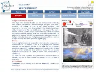
Abteilung Allgemeine Psychologie. Color perception in the intermediate periphery of the visual field. Thorsten Hansen, Lars Pracejus & Karl R. Gegenfurtner. Introduction Perception in the periphery. Contrast sensitivity

E N D
Abteilung Allgemeine Psychologie Color perception in the intermediate periphery of the visual field Thorsten Hansen, Lars Pracejus & Karl R. Gegenfurtner
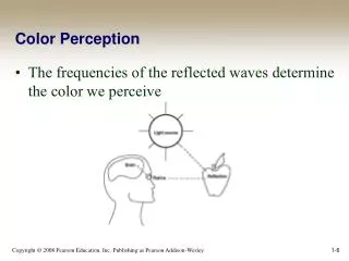
IntroductionPerception in the periphery Contrast sensitivity Fine details (high spatial frequencies) are harder to detect in the periphery. This can be compensated by magnifying the image.
IntroductionCone density in the human retina Curcio et al. (1990) Steep decline towards periphery. Nasal retina has a higher density than temporal retina. Nasal Fovea Temporal 1000 /mm²
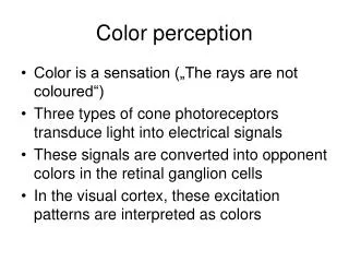
IntroductionColor opponency in the fovea The RF center of a parvo ganglion cell in the fovea is driven by a single cone. Derrington (2001).
IntroductionColor opponency at larger eccentricities …is expected to be weaker and will eventually be zero when the center and surround is driven by an equal ratio of L:M cells Fovea Periphery
IntroductionPsychophysics vs. physiology Macaque ganglion cells remain color sensitive, while human performance drops. Psychophysics (Human) Cell recording (Macaque) Martin et al. (2001). Nature.
L−M Lum S−(L+M) IntroductionPsychophysics »Thus we conclude that there is little or no L/M cone opponent response measurable psychophysically beyond 20–30 deg of eccentricity in the nasal visual field.« Mullen, Sakurai & Chu (2005). Perception.
Methods DKL color space An achromatic axis: Lum Two chromatic axes: L−M and S−(L+M)) Derrington Krauskopf Lennie Lum S−(L+M) L−M S−(L+M) L−M
Methods Procedure Short presentation stimuli (500 ms) forced choice (4AFC) threshold measured by standard stair-case procedure Exp 1 & 2: Detection & Identification Exp. 3: Discrimination
Results Chromatic detection (5 deg stimulus) N = 7 50° 40° 30° 20° 10° in %
Results Identification (5 deg stimulus) N = 7 50° 10° in %
N = 3 L−M Lum S−(L+M) Eccentricity (deg) Results Chromatic detection (8 deg stimulus)
Ellipse was fit to the data to characterize discrimination performance Methods Chromatic discrimination Comparison color varied along 8 chromatic directions
Results Control: Foveal discrimination …as expected: Best at the adaptation point Elongated along the saturation axes Krauskopf & Gegenfurtner (1992). Vision Res.; Hansen, Giesel & Gegenfurtner (in press), J. Vision.
Results Discrimination at 50° Larger size of ellipses: Discrimination is worse, but not absent! Greatest increase along L−M axis Leads to rounder shapes of ellipses off the L−M axis
SummaryChromatic processing in the periphery • Detection • Identification • Discrimination As long as the stimuli are large enough, peripheral color vision is just like foveal vision.
DiscussionSize matters (Mullen et al. 2005) “sinring” stimuli: Radial size was only 1.5 deg.
IntroductionCortical representation Cortical magnification factor The central part of the visual field (10 deg) is represented by about half of all neurons in primary visual cortex V1
IntroductionPhysiology • Size of the receptive fields • Cortical representation • Center – surround ratio • Random wiring • Results physiology – psychophysics
DiscussionBiased sample? (Martin et al. 2001) 34/53 overt red-green response 28/35 cone-opponent 11 not significantly different from foveal cells
Results Cone-opponent thresholds Further evidence for chromatic discrimination
IntroductionColor opponency at larger eccentricities • Several factors can contribute to preserve color opponency • selective wiring (vs. random wiring) • elongated RFs • unequal/random distribution of cone types at each retinal location
IntroductionPhysiology „Random Wiring“ vs. „Selective Wiring“ Jusuf et al., 2006
Introduction Selectivity by elongated RFs Martin et al., 2001 Midget RF centers are elongated and may rotate to sample one cone type more than the other to increase cone-purity
IntroductionElongated RFs can increase the L/(L+M) ratio Martin et al., 2001