Download
1 / 107
1.39k likes | 2.09k Views
Cytogenetics: Karyotypes and Chromosome Aberrations. Chapter 6. 6.1 The Human Chromosome Set. The number and appearance of chromosomes in the nucleus of an organism is an important characteristic Chromosome analysis is a powerful and useful technique in human genetics. Chromosome Number.

E N D
Cytogenetics: Karyotypes and Chromosome Aberrations Chapter 6
6.1 The Human Chromosome Set • The number and appearance of chromosomes in the nucleus of an organism is an important characteristic • Chromosome analysis is a powerful and useful technique in human genetics
Chromosome Number • Chromosome number in selected organisms Table 6-1, p. 121
Chromosome Shape • As chromosomes condense and become visible during cell division, certain structural features can be recognized • Centromere • A region of a chromosome to which microtubule fibers attach during cell division • The location of a centromere gives a chromosome its characteristic shape
Centromere Location • Metacentric • A chromosome that has a centrally placed centromere • Submetacentric • A chromosome whose centromere is placed closer to one end than the other • Acrocentric • A chromosome whose centromere is placed very close to, but not at, one end
Human Chromosomes • Replicated chromosomes at metaphase consist of sister chromatids joined by a single centromere Fig. 6-1, p. 122
Metaphase Chromosomes • Chromosomes are identified by size, centromere location, banding pattern
Metacentric Submetacentric Acrocentric Short arm (p) Satellite p Centromere p Stalk q q Long arm (q) 3 17 21 Fig. 6-2, p. 122
Types of Chromosomes • Sex chromosomes • In humans, the X and Y chromosomes that are involved in sex determination. These have different sizes and shapes • Autosomes • Chromosomes other than the sex chromosomes • In humans, chromosomes 1 to 22 are autosomes
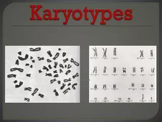
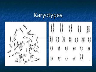
A Set of Human Chromosomes • Human chromosomes are analyzed by construction of karyotypes • Karyotype • A complete set of chromosomes from a cell that has been photographed during cell division and arranged in a standard sequence
A Human Karyotype Fig. 6-3, p. 122
Karyogram: Chromosome Banding Patterns Fig. 6-4, p. 123
1 2 3 4 5 6 7 8 9 10 11 12 13 14 15 16 17 18 19 20 21 22 Y X Fig. 6-4, p. 123
System of Naming Chromosome Bands • Allows any region to be identified by a descriptive address (chromosome number, arm, region, and band)
Band 6 Region 5 3 4 3 2 1 2 2 1 Arm 3 1 2 p 1 1 q 1 2 1 2 3 4 2 5 1 3 2 1 2 4 3 4 1 Fig. 6-5, p. 123
Add a few drops of blood. Add phytohemagglutinin to stimulate mitosis. Draw 10 to 20 ml of blood. Incubate at 37°C for 2 to 3 days. Transfer to tube containing fixative. Transfer cells to tube. Add Colcemid to culture for 1 to 2 hours to stop mitosis in metaphase. Centrifuge to concentrate cells. Add low-salt solution to eliminate red blood cells and swell lymphocytes. Drop cells onto microscope slide. Digitized chromosome images processed to make karyotype. Examine with microscope. Stain slide with Giemsa. Fig. 6-6, p. 124
Metaphase Chromosomes (a) Arranged Into a Karyotype (b) Fig. 6-7, p. 125
Keep In Mind • Karyotype construction and analysis are used to identify chromosome abnormalities
ANIMATION: Preparation of a karyotype To play movie you must be in Slide Show Mode PC Users: Please wait for content to load, then click to play Mac Users: CLICK HERE
6.3 Constructing and Analyzing Karyotypes • Different stains and dyes produce banding patterns specific to each chromosome • Karyotypes reveal variations in chromosomal structure and number • 1959: Discovery that Down syndrome is caused by an extra copy of chromosome 21 • Chromosome banding and other techniques can identify small changes in chromosomal structure
Information Obtained from a Karyotype • Number of chromosomes • Sex chromosome content • Presence or absence of individual chromosomes • Nature and extent of large structural abnormalities
Banding technique Appearance of chromosomes G-banding — Treat metaphase spreads with trypsin, an enzyme that digests part of chromosomal protein. Stain with Giemsa stain. Observe banding pattern with light microscope. Darkly stained G bands. Fig. 6-8a, p. 126
Banding technique Appearance of chromosomes Q-banding — Treat metaphase spreads with the chemical quinacrine mustard. Observe fluorescent banding pattern with a special ultraviolet light microscope. Bright fluorescent bands upon exposure to ultraviolet light; same as darkly stained G bands. Fig. 6-8b, p. 126
Banding technique Appearance of chromosomes R-banding — Heat metaphase spreads at high temperatures to achieve partial denaturation of DNA. Stain with Giemsa stain. Observe with light microscope. Darkly stained R bands correspond to light bands in G-banded chromosomes. Pattern is the reverse of G-banding. Fig. 6-8c, p. 126
Banding technique Appearance of chromosomes C-banding — Chemically treat metaphase spreads to extract DNA from the arms but not the centromeric regions of chromosomes. Stain with Giemsa stain and observe with light microscope. Darkly stained C band centromeric region of the chromosome corresponds to region of constitutive heterochromatin. Fig. 6-8d, p. 126
Chromosomal Aberrations and Specific Syndromes Table 6-2, p. 126
Chromosome Painting • New techniques using fluorescent dyes generate unique patterns for each chromosome Fig. 6-9a, p. 127
Obtaining Cells for Chromosome Studies • Any nucleus can be used to make karyotype • Lymphocytes, skin cells, cells from biopsies, tumor cells • Sampling cells before birth • Amniocentesis • Chorionic villus sampling (CVS)
Amniocentesis • A method of sampling the fluid surrounding the developing fetus by inserting a hollow needle and withdrawing suspended fetal cells and fluid • Used in diagnosing fetal genetic and developmental disorders • Usually performed in the sixteenth week of pregnancy
Removal of about 20 ml of amniotic fluid containing suspended cells that were sloughed off from the fetus A few biochemical analyses with some of the amniotic fluid Centrifugation Quick determination of fetal sex and analysis of purified DNA Fetal cells Growth for several days in culture medium Biochemical analysis for the presence of alleles that cause many different metabolic disorders Karyotype analysis (a) Fig. 6-10a, p. 127
Chorionic Villus Sampling (CVS) • A method of sampling fetal chorionic cells by inserting a catheter through the vagina or abdominal wall into the uterus • Used in diagnosing biochemical and cytogenetic defects in the embryo • Usually performed in the eighth or ninth week of pregnancy
Chorionic villi Developing placenta Ultrasound to monitor procedure Developing fetus Bladder Uterus Chorion Catheter Amniotic cavity Rectum (a) Fig. 6-11a, p. 128
Exploring Genetics: Noninvasive Prenatal Diagnosis • Methods are being investigated to isolate fetal cells that can pass into the mother’s bloodstream (placental cells, white blood cells, immature red blood cells) for genetic testing p. 129
ANIMATION: Amniocentesis To play movie you must be in Slide Show Mode PC Users: Please wait for content to load, then click to play Mac Users: CLICK HERE
6.4 Variations in Chromosome Number • Changes in chromosome number or chromosome structure can cause genetic disorders • Two major types of chromosomal changes can be detected in a karyotype • A change in chromosomal number • A change in chromosomal arrangement
Changes in Chromosome Number • Polyploidy • A chromosomal number that is a multiple (3n or 4n) of the normal haploid chromosomal number • Aneuploidy • A chromosomal number that is not an exact multiple of the haploid number
Polyploidy Changes the Number of Chromosome Sets • Triploidy • A chromosomal number that is three times the haploid number, having three copies of all autosomes and three sex chromosomes • Tetraploidy • A chromosomal number that is four times the haploid number, having four copies of all autosomes and four sex chromosomes
A Triploid Karyotype Fig. 6-12, p. 130
A Triploid Infant Fig. 6-13, p. 131
Keep In Mind • Polyploidy results when there are more than two complete sets of chromosomes
Aneuploidy Involves the Gain or Loss of Individual Chromosomes • Monosomy • A condition in which one member of a chromosomal pair is missing; one less than the diploid number (2n – 1) • Trisomy • A condition in which one chromosome is present in three copies, and all others are diploid; one more than the diploid number (2n + 1)
Causes of Aneuploidy • Nondisjunction • The failure of homologous chromosomes to separate properly during meiosis
Extra chromosome (n + 1) Nondisjunction Extra chromosome (n + 1) Missing chromosome (n − 1) Missing chromosome (n − 1) Meiosis I Meiosis II Gametes (a) Fig. 6-14a, p. 132
Nondisjunction Extra chromosome (n + 1) Normal division Missing chromosome (n− 1) Normal (n) Normal (n) Meiosis I Meiosis II Gametes (b) Fig. 6-14b, p. 132
Effects of Monosomy and Trisomy • Autosomal monosomy is a lethal condition • Eliminated early in development (spontaneous abortion) • Some autosomal trisomies are relatively common • Most result in spontaneous abortion • Three types can result in live births (13, 18, 21)