Download
1 / 20
200 likes | 401 Views
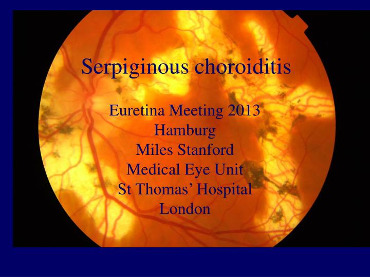
Serpiginous choroiditis. Euretina Meeting 2013 Hamburg Miles Stanford Medical Eye Unit St Thomas’ Hospital London. Serpiginous choroiditis. Rare Bilateral 40-60 years Mainly caucasian Slight preponderance for men. Serpiginous choroiditis - pathology. Little available

E N D
Serpiginouschoroiditis Euretina Meeting 2013 Hamburg Miles Stanford Medical Eye Unit St Thomas’ Hospital London
Serpiginous choroiditis • Rare • Bilateral • 40-60 years • Mainly caucasian • Slight preponderance for men
Serpiginous choroiditis - pathology • Little available • Widespread atrophy of photoreceptors, RPE and choriocapillaris • Lymphocytic infiltration of the choroid • Secondary choroidal neovascularisation
Serpiginous choroiditis – clinical features • Unilateral decrease in central vision, metamorphopsia or scotoma • Little anterior segment reaction • Lesions classically peripapillary and then spread outwards • Disease progression is stepwise and asymmetric • Eventually permanent scar and subretinal fibrosis
Serpiginous choroiditis – stepwise progression over 18 months
Fluorescein angiography showing early masking and late staining on the edge of a old scar
Serpiginous – FFA staining at the edge of an old scar. These changes may be more evident on ICG
Serpiginous choroiditisdifferential diagnosis • APMPPE • Myopia • Choroidal ischaemia • Sarcoidosis • Toxoplasma • Tuberculosis/syphilis • Metastases/lymphoma • Retinochoroidal dystrophies
Ampiginous choroiditis (mantoux 20mm, subsequently developed Eales’ disease)
Serpiginous-like choroiditis and TB • Presumed uveitis due to TB: All patients with 1 year follow up, exclusion of other infections, +ve Mantoux, no recurrence after full anti TB treatment • 26/192 (15%) patients with presumed TB-related posterior uveitis had serpiginous like choroiditis (OR 26; 95% CI 7.4-91.4) • Sensitivity 14%: specificity 98%: positive predictive value 56% • Not a good sign for screening but makes diagnosis 90% certain if positive Gupta A et al Am J Ophthalmol 2010 149:562
Serpiginous-like choroiditis and TB • 11/21 (52%) patients tested +ve with Quantiferon compared to 9% HC and 13% uveitis controls • 3/11 improved with specific anti-TB treatment • ?directly due to bacteria or allergic response Mackensen F et al Am J Ophthalmol 2008 146;761
Serpiginous-like choroiditis and TB • Comparison of 5 patients with serpiginous like (SLC) and classical serpiginous (SC) • Patients with SLC were: - most likely to have come from a country where TB endemic - To have unilateral multifocal disease with significant vitritis - to have a positive PPD - to respond to tuberculostatic therapy Arch Ophthalmol 2010 128: 853
Serpiginous choroiditisInvestigations • FFA • ICG • OCT • Electrodiagnostics • Visual fields • Mantoux/IFN gamma
Serpiginous choroiditis - complications • CNVM occurs in 15-35% • Usually arises from the edge of a scar but may be peripapillary • Serous retinal or RPE detachments • Subretinal fibrosis • Rarely, CMO or NVs
Serpiginous choroiditisTreatment • Goals of therapy are to control active lesions rapidly and to prevent further recurrences and progression • Steroids – oral or pulsed • Other immunosuppressives • Infliximab • Treatment for secondary neovascularisation
Serpiginous choroiditis - prognosis • Very few long term studies • Chronic, progressive disease in a stepwise manner • Active lesions usually resolve over 3-6 months but may take longer • Extrafoveal lesions may not give rise to symptoms and so pass unrecognised
Serpiginous choroiditis - Conclusions • Rare, progressive disease of the middle-aged • Must exclude TB especially if patient from endemic area • Treat with standard immunosuppressives to control active lesions and prevent progression • Potential for secondary CNVM