Download
1 / 27
270 likes | 480 Views
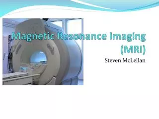
Magnetism & MRI (difference between magnetic & MRI ) Biophysics (PHR 177). Prof. Dr. Moustafa . M. Mohamed Vice Dean Dr. Eng. Safa Ahmed El- Askary Faculty of Allied Medical Science Pharos University Alexandria. M -AGNETIC R -ESONANCE I -MAGING.

E N D
Magnetism & MRI (difference between magnetic & MRI ) Biophysics (PHR 177) Prof. Dr. Moustafa. M. Mohamed Vice Dean Dr. Eng. Safa Ahmed El-Askary Faculty of Allied Medical Science Pharos University Alexandria
Physics of MRIIt is an interplay of • Magnetism • Resonance
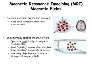
TYPES OF ATOMIC MOTION&LAW OF ELECTROMAGNETISM • The electron orbits the nucleus • The electron spins on its own axis • ***The nucleus spins on its own axis***. • A charged particle in motion will create a magnetic field
MRI USES THE HYDROGEN ATOM • The nucleus contains no neutrons but contains 1 proton • THE HYDROGEN NUCLEUS HAS A NET POSITIVE CHARGE • Hydrogen nucleus is a spinning, positively charged particle. • Very abundant in the human body-H20 • Has a large magnetic moment
Introduction • What is MRI? • Magnetic resonance imaging (MRI) is a spectroscopic imaging technique used in medical settings to produce images of the inside of the human body. • MRI is based on the principles of nuclear magnetic resonance (NMR), which is a spectroscopic technique used to obtain microscopic chemical and physical data about molecules • In 1977 the first MRI exam was performed on a human being. It took 5 hours to produce one image.
Introduction • How Does it Work? • The magnetic resonance imaging is accomplished through the absorption and emission of energy of the radio frequency (RF) range of the electromagnetic spectrum.
The Components: • A magnet which produces a very powerful uniform magnetic field. • Gradient Magnets which are much lower in strength and are used to create a variable field. 3. Equipment to transmit radio frequency (RF). 4. A very powerful computer system, which translates the signals transmitted by the coils. • The magnets currently used in scanners today are in the .5-tesla to 2.0-tesla range (5,000 to 20,000-gauss). Earth magnetic field: 0.5-gauss
The Technology • How Does It All Work? • Spin: • The atoms that compose the human body have a property known as spin(a fundamental property of all atoms in nature like mass or charge). • Spin can be thought of as a small magnetic field and can be given a + or – sign . • Components of an atom such as protons, electrons and neutrons all have spin.
PRECESSION Precessional path • Due to the influence of B0, the hydrogen nucleus “wobbles” or precesses (like a spinning top as it comes to rest) • The axis of the nucleus forms a path around B0 known as the “precessional path” B0 Hydrogen nucleus
PRECESSION • The speed at which hydrogen precesses depends on the strength of B0 and is termed the “precessional frequency” • The precessional frequency of hydrogen in a 1.5 Tesla magnetic field is 63.86 MHz • The precessional paths of the individual hydrogen nucleus’ is random, or “out of phase”
The Technology (cont.) • Human body is mainly composed of fat and water, which makes the human body composed of about 63% hydrogen. • Why Are Protons Important to MRI? • positively charged • spin about a central axis • a moving (spinning) charge creates a magnetic field. • the straight arrow (vector) indicates the direction of the magnetic field.
Fig: 1. A) The top spinning in the earth's gravity. The gravity tries to pull it down but it stays upright due to its fast rotation. B) A charge spinning in the magnetic field Bo.
The Technology (cont.) • When placed in a large magnetic field, hydrogen atoms have a strong tendency to align in the direction of the magnetic filed • Inside the bore of the scanner, the magnetic field runs down the center of the tube in which the patient is placed, so the hydrogen protons will line up in either the direction of the feet or the head. • The majority will cancel each other, but the net number of protons is sufficient to produce an image.
NET MAGNETIZATION VECTOR • An excess of hydrogen nuclei will line up parallel to B0 and create the NMV of the patient
The Technology (cont.) • Energy Absorption: • The MRI machine applies radio frequency (RF) pulse that is specific to hydrogen. • The RF pulses are applied through a coil that is specific to the part of the body being scanned.
The Technology (Cont.) Resonance (cont.) The gradient magnets are rapidly turned on and off which alters the main magnetic field. • The pulse (RF) directed to a specific area of the body causes the protons to absorb energy and spin in different direction, which is known as Resonance Frequency (Hz) of energyabsorption depends on strength of external magnetic field.
Fig: 2 A) The compass needle (a small magnet) aligns itself with a N/S-S/N direction when placed in a large magnetic field. B) When another strong magnet is brought near the aligned compass needle the magnetic fields of all three magnets interact in such a way that the mobile, weakest magnet (the compass needle) realigns itself away from its original orientation. C) When the perturbing magnetic field is removed suddenly the compass needle magnet realigns itself with the large external magnet field, but before realigning, it wobbles around the point of stability and gradually comes to rest.
The Technology (cont.) • The resonance frequency, 0, is referred to as the Larmor frequency.
The Technology (cont.) • Imaging: • When the RF pulse is turned off the hydrogen protons slowly return to their natural alignment within the magnetic field and release their excess stored energy. This is known as relaxation. • What happens to the released energy? • Released as heat OR • Exchanged and absorbed by other protons OR • Released as Radio Waves.
A CHANGING MAGNETIC FIELD IS CREATED IF YOU PLACE A RECEIVER COIL (ANTENNA) IN THE PATH OF THE CHANGING MAGNETIC FIELD, A CURRENT WILL BE INDUCED THIS IS FARADAY’S LAW OF INDUCTION
FARADAY’S LAW OF INDUCTION A changing magnetic field will induce an electrical current in any conducting medium COILS • Used to: • transmit pulses of radiofrequency energy • receive induced voltage - MR SIGNAL • increase image quality by tuning in to one body part at a time
MR SIGNAL • Collected by a coil • Encoded through a series of complex techniques and calculations (magic?) • Stored as data • Mapped onto an image matrix
ADVANTAGES • Superior soft tissue contrast resolution - excellent pathological discrimination • No ionizing radiation • Direct multi-planar imaging (transverse, coronal, sagittal, any oblique) • Non-invasive .