Download
1 / 18
180 likes | 246 Views
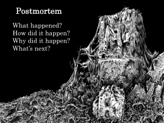
The Role of Agonal Factors in Human Postmortem CNS Research. Daniel W. McKeel, Jr., M.D. ADRC Neuropathology Tissue Resource Washington U. School of Medicine St. Louis MO - 6/23/04. MEDLINE Literature Review. Category 4 citations were examined in detail for factors

E N D
The Role of Agonal Factors in Human Postmortem CNS Research Daniel W. McKeel, Jr., M.D. ADRC Neuropathology Tissue Resource Washington U. School of Medicine St. Louis MO - 6/23/04
MEDLINE Literature Review Category 4 citations were examined in detail for factors pertinent to using ADRC postmortem brain material for biochemical research
Coma, MOF, respi-ratory arrest, hypox. Brain pH (need a standard method!) PMI Febrile state Terminal medications Age and Gender Brain lobe (regional) mRNA heterogeneous factor effects add to varying stability on yield and quality Gene expression & postmortem CNS pH: Low: depressed energy, proteolysis High: elevated stress, transcription factors Identified Agonal Factors
Li JZ, Vawter MP, Walsh DM, Tomita H, Evans SJ, Choudary PV, Lopez JF, Avelar A, Shokoohi V, Chung T, Mesarwi O, Jones EG, Watson SJ, Akil H, Bunney WE Jr, Myers RM. Systematic changes in gene expression in postmortem human brains associated with tissue pH and terminal medical conditions. [Journal Article] Human Molecular Genetics. 13(6):609-16, 2004 March 15 • We observed a remarkable degree of natural variation among 120 samples, which represented three brain regions in 40 subjects. • Individuals who suffered prolonged agonal states, such as with respiratory arrest, multi-organ failure or coma, tended to have lower pH in the brain • Those who experienced brief deaths, associated with accidents, cardiac events or asphyxia, generally had normal pH. • The lower pH samples exhibited a systematic decrease in expression of genes involved in energy metabolism and proteolytic activities, and a consistent increase of genes encoding stress-response proteins and transcription factors.
Tomita H, Vawter MP, Walsh DM, Evans SJ, Choudary PV, Li J, Overman KM, Atz ME, Myers RM, Jones EG, Watson SJ, Akil H. Bunney WE Jr. Effect of agonal and postmortem factors on gene expression profile: quality control in microarray analyses of postmortem human brain.Biological Psychiatry. 55(4):346-52, 2004 • Coma and hypoxia do affect RNA integrity and gene expression profiles more than age, gender & postmortem factors. Propose “Average Correlation Index” to reduce specimen variability.
Many factors affect mRNA • Preece P, Cairns NJ. Quantifying mRNA in postmortem human brain: influence of gender, age at death, postmortem interval, brain pH, agonal state and inter-lobe mRNA variance. [Journal Article] Brain Research. Molecular Brain Research. 118(1-2):60-71, 2003 Oct 21 • TaqMan RT-PCR measured 7 mRNAs • 90 AD and 81 control brains (lobar mRNA same) • Females had less mRNA than males • Brain pH & amount of RNA (+) corr. except GFAP • “Agonal state a poor predictor of mRNA levels”
Cummings TJ, Strum JC, Yoon LW, Szymanski MH, Hulette CM. Recovery and expression of messenger RNA from postmortem human brain tissue.Modern Pathology. 14(11):1157-61, 2001 Nov • Bryan ADRC Rapid Autopsy Program at Duke • 10 AD + 9 Controls (1 to 11 hr PMI) • 19 brains RNA integrity + mRNA gene expression (CSF pH, fever/sepsis, O2, sudden?) • “All samples yield intact RNA without degradation” (“successful gene expression may require enhanced procurement efforts”)
Bissette G, Seidler FJ, Nemeroff CB, Slotkin TA. High affinity choline transporter status in Alzheimer's disease tissue from rapid autopsy. [Review] [12 refs] [Review] Annals of the New York Academy of Sciences. 777:197-204, 1996 • Choline transporter degrades rapidly • Brains acquired within 2 hours of death • Choline transporter increased in AD cortex compared to non-AD controls • Putamen used as a “spared” control region
What Can NACC/NIA Do? [1] • Advertise its frozen brain resources! • Broker tissue distribution requests that involve multiple ADRCs (clearing house) • Encourage collaborative grants & symposia to standardize frozen tissue collection methods • Explore feasibility of regional specialized brain banks (genomics, proteomics, laser capture microdissection analysis of single and pooled cells) -- rigid acceptance criteria for specimens
What Can NACC/NIA Do? [2] • Gather and distribute center-specific specimen requests and distribution data! • Tabulate center-specific practical experience with using agonal factor data to facilitate research. • Agonal factor use and outcome research within the ADRC community - what factors matter? • Add agonal factors to the NACC database and make this data widely available to investigators
What Can ADRC Pathologists Do? • Use existing brain banking protocols to formulate a standard protocol for all centers. • Develop standard tissue block label protocols to facilitate collaborative ADRC research • Adopt the McKeel-Gado visual stds-based system (Brain Pathol 1994) for scoring brain atrophy and ventricular dilatation at autopsy. • Develop standard CSF collection protocols • Add banked CNS/CSF requests received and fulfilled to the NACC-reportable data
Frozen Human Brain Protocols • Nochlin D et al. (U. Washington), Acta Neuropathologica. 86(6):645-50, 1993 • Aluminum plates chilled with dry ice (CO2) • CNS suitable for LM, EM + biochemistry • Vonsattel, McKee, Hedley-Whyte et al. (Harvard), JNeuropathol Experimental Neurology. 54(1):42-56, 1995 • Aluminum plates chilled with dry ice (CO2) • Top plate to flatten specimen (coronal slices) • Standardized block sampling protocol
Scoring CNS Atrophy & Ventricular Size at Autopsy McKeel DW Jr, Gado M. A visual standards based system for scoring Alzheimer and aging-related human brain atrophy at autopsy (abstr. P34-11).Brain Pathol 1994;4:544
WUSM ADRC Standard Blocks 1. Frontal cortex 2. STG + MTG 3. Inf. Parietal ctx 4. Primary visual 5. Hippocampus/ERC ten levels 6. Striatum 7. Mamillary bodies 8. Thalamus 9. Nigra, rostral 10. Nigra, caudal 11. Pons, 3 levels 12. Medulla, 2 levels 13. Spinal Cord 14. Cbellum + Dent. N. 15. Cbellar vermis 16. Hypothalamus 17. Nucleus basalis 18. Orbitofrontal ctx 19. Ant. Cingulate 20. Inf. Temporal ctx 21. Primary motor ctx 22. Primary sensory ctx 23. Amygdala 24. Olfact. Tract & Bulb & ant. olf. nucleus 25. Optic chiasm & nerve 26. WM, deep frontal 27. WM, mid portion 28. WM, occipital 29. Caudate, putamen & globus pallidus 30. Posterior cingulate 31ff - Pathologic lesions
Standardized Immunohistochemistry • At present no standardization exists in IHC methodology among ADRCs • Includes fixation, embedding materials, pretreatment protocols, reagent sources, antibody working titers, substrates used, etc. • Hence results vary non-systematically and adversely affect comparisons among results obtained at various centers.
CDR 0 Hipp:10D5 Aß+ PHF1 Dual IHC Braak & Braak Neurofibrillary STAGE III
CDR 3 AD Hipp:10D5 Aß + PHF1 Braak Stage VI
There is lots of Work to do! Standardization now will yield major dividends in the future.