Download
1 / 14
160 likes | 479 Views
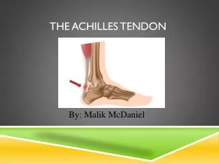
Peroneal and Achilles tendon problems with surgical management. Peroneal tendon stabilisation. Anatomy. Peroneal tendons course behind the distal fibula The peroneus brevis may have degenerative changes if the injury is not identified in a timely fashion. What happens and what you see.

E N D
Peroneal and Achilles tendon problems with surgical management
Anatomy Peroneal tendons course behind the distal fibula The peroneusbrevis may have degenerative changes if the injury is not identified in a timely fashion
What happens and what you see The peronealretinaculum may be avulsed from the fibula or calcaneus or lifted up enough to allow tendon dislocation Forceful contraction of peroneals during sudden dorsiflexion and inversion
Classification Figure 26-65 Classification of pathology in peroneal tendon dislocations. A, Normal. B, Grade I: superior peronealretinaculum stripped off fibula. C, Grade II: fibrous rim avulsed from the posterolateral aspect of the fibula along with the superior peronealretinaculum. D, Grade III: bony avulsion of the posterolateral part of the fibula by the superior peronealretinaculum. (Modified from Eckert WR, Davis EA Jr: J Bone Joint Surg Am 58:670-673, 1976.)
Management 1 Direct repair • If acute repair correctly • Anchors useful to aid stabilisation of retinaculum
Management 2 Groove deepening repair Split the fibula distally leaving a posterior hinge intact, by curetting out some of the cancellous bone and replacing the hinge the groove will be deepened Like trochleoplasty of the knee allowing a deeper grove for the tendons to sit in, minimising further dislocation
Management 3 DuVries Bone block lateral fibula osteotomy procedure Modification of Kelly procedure Creates new groove for tendons to sit in, posterior to the fibula
Approach to Achilles/Haglunds • Can approach medially or laterally • If lateral find and protect the sural nerve • Try and avoid central/splitting incision
Haglunds decompression I • Try to remove degenerative tissue and decrease impingement • Can take away more than you think
Haglunds decompression II Can resect/decompress with osteotome or burr. If involving greater than 50% of achillesstabilsie with anchors
FHL transfer • Severe unremitting insertionaltendinopathy • Excessive calcificinsertionaltendinopathy • Failed anchors resulting in pullout
Principles of FHL transfer 2 incisions: Medial over Achilles and Medial over knot of Henry Deliver FHL into Achilles wound and secure to OS Calsis with biotenodesis screw at appropriate tension NB don’t forget to tenodese distal FHL with FDL to enable flexion at big toe