Download
1 / 41
420 likes | 550 Views
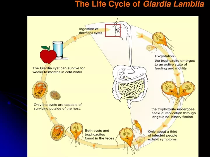
The Life Cycle of Giardia Lamblia. Giardia lamblia The cysts, are oval measuring from 11 to 14 µm in length and 7 to 10 µm in width. Four nuclei are usually visible along with axonemes, and median bodies. Often, the cyst will appear shrunken, pulling away from the cyst wall.

E N D
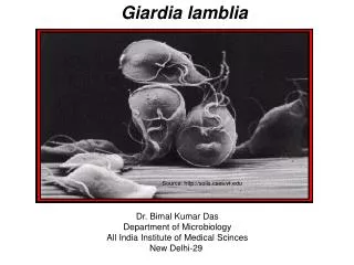
Giardia lambliaThe cysts, are oval measuring from 11 to 14 µm in length and 7 to 10 µm in width. Four nuclei are usually visible along with axonemes, and median bodies. Often, the cyst will appear shrunken, pulling away from the cyst wall
Giardia lambliaThe trophozoites are described as having a 'tear-drop' shape and are 18 µm long and 10 µm wide. The trophozoites contain two nuclei, four pair of flagella, two axonemes, and two curved bodies called the median bodies.
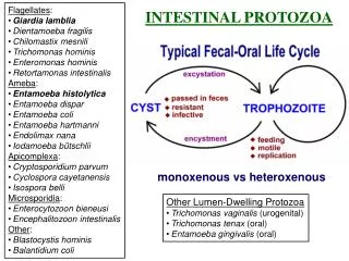
Entamoeba histolytica Life cycle the cysts can survive days to weeks in the external environment and are responsible for transmission Trophozoites passed in the stool are rapidly destroyed once outside the body
Entamoeba histolytica cyst will have 1 to 4 nuclei. In addition, the peripheral chromatin is generally evenly distributed. trophozoite15 to 20 µm. CYST 10 – 15 µM
Ascariasis is a disease caused by Ascaris Lumbricoides, an intestinal roundworm known as a nematode
Ascaris lumbricoides life cycle Female 50 cm Male 30 cm
Fertilized, corticated eggsAscaris AscarisFertilized, decorticated eggs size 45 - 85 microns
Ascarisunfertilized or otherwise non-viable eggs Egg size(infertile): 78 - 105 micronsin length Both fertile and infertile ova can be corticated or smooth
The life cycle of Enterobius vermicularis Female 8-13mm Male 2-5mm
Eggs of the human pinworm Enterobius vermicularis Egg size 50-60 µm by 20-32 µm
Strongyloides stercoralis life cycle Egg size: 52 - 58 microns Rhabditiform larva length: 250 microns Filariform larva: 550 microns 3 or 4days to filariform 2-5 days
Hookworm Ancylostoma braziliense] [Ancylostoma caninum] [Ancylostoma duodenale] [Necator americanus] infective larvae can survive 3 to 4 weeks in favorable environmental conditions after 5 to 10 days larvae hatch in 1 to 2 days.
Necator americanus : 5 to 11 millimetres , 90 percent of human hookworm infections that occur in tropical and subtropical regions of the world Ancylostoma duodenale, 8 to 13 millimeters long, is found on all continents but is most prevalent in warm regions. A. braziliense, from 8 to 11 millimetres long, is normally parasitic in dogs and cats; man, however, is sometimes infected by this species in the southern United States, South America, and Asia.
Hookworm filariform larva Rhabditiform larvae that hatch from eggs are 250-300 µm long and approximately 15-20 µm wide.
Hookworm eggs in unstained wet mounts, taken at 400× magnification 60-75 µm by 35-40 µm.
Ancylostoma duodenale Copulatory bursa
Plasmodium Anopheles mosquito
Recognizing Erythrocytic Stages:Schematic Morphology TROPHOZOITE RING SCHIZONT GAMETOCYTE
Plasmodium falciparum Infected erythrocytes: normal size M I Gametocytes: mature (M)and immature (I) forms (I is rarely seen in peripheral blood) Rings: double chromatin dots; appliqué forms; multiple infections in same red cell Schizonts: 8-24 merozoites (rarely seen in peripheral blood) Trophozoites: compact (rarely seen in peripheral blood)
Plasmodium vivax Infected erythrocytes: enlarged up to 2X; deformed; (Schüffner’s dots) Rings Trophozoites: ameboid; deforms the erythrocyte Schizonts: 12-24 merozoites Gametocytes: round-oval
Plasmodium ovale Infected erythrocytes: moderately enlarged (11/4 X); fimbriated; oval; (Schüffner’s dots) “malariae - like parasite in vivax - like erythrocyte” Trophozoites: compact Rings Gametocytes: round-oval Schizonts: 6-14 merozoites; dark pigment; (“rosettes”)
Plasmodium malariae • Infected erythrocytes: size normal to decreased (3/4X) Trophozoite: typical band form Trophozoite: compact Schizont: 6-12 merozoites; coarse, dark pigment Gametocyte: round; coarse, dark pigment
Morphology • Malarial parasite trophozoites are generally ring shaped, 1-2 microns in size, although other forms (ameboid and band) may also exist. • The sexual forms of the parasite (gametocytes) are much larger and 7-14 microns in size. • P. falciparum is the largest and is banana shaped, while others are smaller and round.
Morphology Ring form: (Plasmodium vivax ) a ring of bluish cytoplasm with a dot-like nucleus
Trophzoite of Plasmodium vivax • irregular cytoplasm and enlarged nucleus with malarial pigment • ( hemozoin)
Schizont of P.vivax • multiple masses of nuclear chromatin
Gametocytes Male gametocyte Female gametocyte Note: compact cytoplasm and absence of nuclear division.
Ring form of P. falciparum Ring with double nuclei Multiple infections
African sleep sickness Trypanosoma brucei gambiense Tsetse Fly
Trypanosoma Diagnosis 1- Detection of trypanosoma chancre after bite African Trypanosomiasis in Travelers Returning to the United Kingdom The Tsetse bite is usually painful and may develop into a red sore called a chancre
2- Blood smear within 21 days from the bit , it will show the parasite A blood smear from a patient with African trypanosomiasis. a centrally located nucleus, an undulating membrane, and an anterior flagellum. The two Trypanosoma brucei species that cause human trypanosomiasis, T. b. gambiense and T. b. rhodesiense, are undistinguishable morphologically. The trypanosomes length range is 14-33 µm
Trypanosoma brucei gambiense • trypomastigote