Download
1 / 1
10 likes | 240 Views
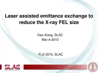
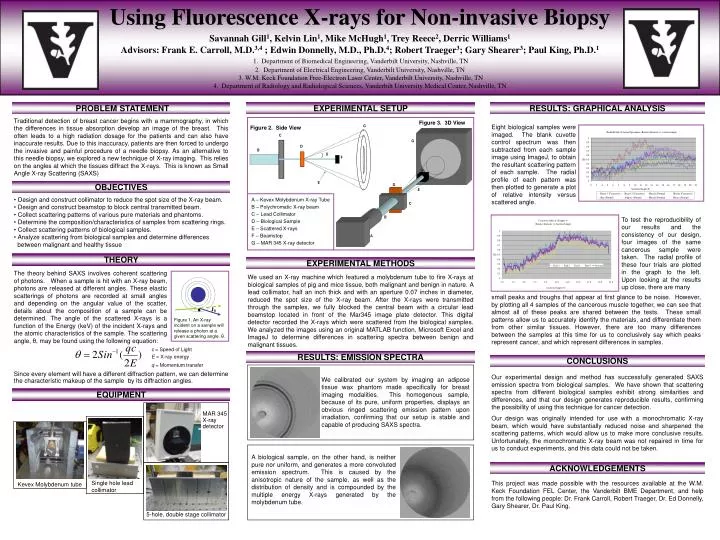
θ. G. C. G. D. B. θ. F. F. D. E. E. A – Kevex Molybdenum X-ray Tube B – Polychromatic X-ray beam C – Lead Collimator D – Biological Sample E – Scattered X-rays F – Beamstop G – MAR 345 X-ray detector. C. B. A.

E N D
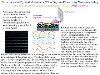
θ G C G D B θ F F D E E A – Kevex Molybdenum X-ray Tube B – Polychromatic X-ray beam C – Lead Collimator D – Biological Sample E – Scattered X-rays F – Beamstop G – MAR 345 X-ray detector C B A Figure 1. An X-ray incident on a sample will release a photon at a given scattering angle, θ. MAR 345 X-ray detector Kevex Molybdenum tube Single hole lead collimator 5-hole, double stage collimator Using Fluorescence X-rays for Non-invasive Biopsy Savannah Gill1, Kelvin Lin1, Mike McHugh1, Trey Reece2, Derric Williams1 Advisors: Frank E. Carroll, M.D.3,4 ; Edwin Donnelly, M.D., Ph.D.4; Robert Traeger3; Gary Shearer3; Paul King, Ph.D.1 1. Department of Biomedical Engineering, Vanderbilt University, Nashville, TN 2. Department of Electrical Engineering, Vanderbilt University, Nashville, TN 3. W.M. Keck Foundation Free-Electron Laser Center, Vanderbilt University, Nashville, TN 4. Department of Radiology and Radiological Sciences, Vanderbilt University Medical Center, Nashville, TN PROBLEM STATEMENT EXPERIMENTAL SETUP RESULTS: GRAPHICAL ANALYSIS Traditional detection of breast cancer begins with a mammography, in which the differences in tissue absorption develop an image of the breast. This often leads to a high radiation dosage for the patients and can also have inaccurate results. Due to this inaccuracy, patients are then forced to undergo the invasive and painful procedure of a needle biopsy. As an alternative to this needle biopsy, we explored a new technique of X-ray imaging. This relies on the angles at which the tissues diffract the X-rays. This is known as Small Angle X-ray Scattering (SAXS) Figure 3. 3D View Eight biological samples were imaged. The blank cuvette control spectrum was then subtracted from each sample image using ImageJ, to obtain the resultant scattering pattern of each sample. The radial profile of each pattern was then plotted to generate a plot of relative intensity versus scattered angle. Figure 2. Side View OBJECTIVES • Design and construct collimator to reduce the spot size of the X-ray beam. • Design and construct beamstop to block central transmitted beam. • Collect scattering patterns of various pure materials and phantoms. • Determine the composition/characteristics of samples from scattering rings. • Collect scattering patterns of biological samples. • Analyze scattering from biological samples and determine differences • between malignant and healthy tissue To test the reproducibility of our results and the consistency of our design, four images of the same cancerous sample were taken. The radial profile of these four trials are plotted in the graph to the left. Upon looking at the results up close, there are many THEORY EXPERIMENTAL METHODS The theory behind SAXS involves coherent scattering of photons. When a sample is hit with an X-ray beam, photons are released at different angles. These elastic scatterings of photons are recorded at small angles and depending on the angular value of the scatter, details about the composition of a sample can be determined. The angle of the scattered X-rays is a function of the Energy (keV) of the incident X-rays and the atomic characteristics of the sample. The scattering angle, θ, may be found using the following equation: We used an X-ray machine which featured a molybdenum tube to fire X-rays at biological samples of pig and mice tissue, both malignant and benign in nature. A lead collimator, half an inch thick and with an aperture 0.07 inches in diameter, reduced the spot size of the X-ray beam. After the X-rays were transmitted through the samples, we fully blocked the central beam with a circular lead beamstop located in front of the Mar345 image plate detector. This digital detector recorded the X-rays which were scattered from the biological samples. We analyzed the images using an original MATLAB function, Microsoft Excel and ImageJ to determine differences in scattering spectra between benign and malignant tissues. small peaks and troughs that appear at first glance to be noise. However, by plotting all 4 samples of the cancerous muscle together, we can see that almost all of these peaks are shared between the tests. These small patterns allow us to accurately identify the materials, and differentiate them from other similar tissues. However, there are too many differences between the samples at this time for us to conclusively say which peaks represent cancer, and which represent differences in samples. c = Speed of Light E = X-ray energy q = Momentum transfer RESULTS: EMISSION SPECTRA CONCLUSIONS Since every element will have a different diffraction pattern, we can determine the characteristic makeup of the sample by its diffraction angles. Our experimental design and method has successfully generated SAXS emission spectra from biological samples. We have shown that scattering spectra from different biological samples exhibit strong similarities and differences, and that our design generates reproducible results, confirming the possibility of using this technique for cancer detection. Our design was originally intended for use with a monochromatic X-ray beam, which would have substantially reduced noise and sharpened the scattering patterns, which would allow us to make more conclusive results. Unfortunately, the monochromatic X-ray beam was not repaired in time for us to conduct experiments, and this data could not be taken. We calibrated our system by imaging an adipose tissue wax phantom made specifically for breast imaging modalities. This homogenous sample, because of its pure, uniform properties, displays an obvious ringed scattering emission pattern upon irradiation, confirming that our setup is stable and capable of producing SAXS spectra. EQUIPMENT A biological sample, on the other hand, is neither pure nor uniform, and generates a more convoluted emission spectrum. This is caused by the anisotropic nature of the sample, as well as the distribution of density and is compounded by the multiple energy X-rays generated by the molybdenum tube. ACKNOWLEDGEMENTS This project was made possible with the resources available at the W.M. Keck Foundation FEL Center, the Vanderbilt BME Department, and help from the following people: Dr. Frank Carroll, Robert Traeger, Dr. Ed Donnelly, Gary Shearer, Dr. Paul King.