Download

1 / 63
630 likes | 759 Views
GI Revision. J Rose. Revision is seeing what you have already seen but with some hope of understanding it this time.

E N D
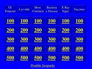
GI Revision J Rose
Revision is seeing what you have already seen but with some hope of understanding it this time. • A revision lecture is all the other lectures shoved together so that the jokes are squeezed out.
Lecture Plan • DYSPHAGIA • JAUNDICE • DIARRHOEA • CROHN’S DISEASE
Dysphagia No patient ever complained of ‘dysphagia’
What does the patient mean? • Can’t chew properly (mastication) • Can’t get it over (deglutition) • After swallowing gets stuck (obstructive dysphagia) or causes pain (odynophagia) • Feels full after eating (bloating or satiety)
Can’t chew properly • Poor teeth, loose dentures, sore gums • Dry mouth • Drugs • Sjögren’s • Weak muscles • Myasthenia - myopathy - dystrophy
Can’t get it over Neuromuscular Incoordination • Cerebrovascular Disease • Motor Neurone Disease • Muscular dystrophy (e.g. myotonic) • Myasthenia gravis • Pouches, webs, high strictures - Skin disease
Gets stuck • Site indicated by time to sticking • Proximal examination inadequate • Sticks and goes down < sticks and comes up • Progression worrying • Liquids easier than solids • Stricture • Peptic • Tumour – Intrinsic or Extrinsic – Ca bronchus • Connective tissue disease • CREST/Systemic sclerosis • Solids and liquids equally affected from the start • Dysmotility • Persistent - Achalasia • Intermittent – Tertiary contractions, presbyoesophagus, nutcracker oesophagus etc.
Just Hurts Oesophagitis • Candida • Usually painless. Check under dentures. Angular cheilitis. Steroid inhalers. • Herpes, CMV • Immunosuppressed. • Opportunistic • AIDS • Reflux • Acid and bile • Iatrogenic • Tubes, ulcerogenic drugs, anticholinergics • Allergic • Eosinophilic Ulceration • Malignant • Adeno- & squamous carcinoma • Extrinsic mediastinal
Investigations • Deglutition problems • Videofluoroscopy with SALT • Obstructive dysphagia or odynophagia • Upper GI endoscopy +/- biopsy • Dysmotility • Barium Meal +/- bread or marshmallow barium bolus • Oesophageal manometry
Axioms • If the sclerae aren’t yellow it’s not jaundice • Carotinaemia? • detect at around 50micromol/l • If there’s no bile in the urine it’s not a hepatobiliary problem • Haemolysis • Gilbert’s • Other metabolic disordersof conjugation • Jaundice with stigmata of CLD is usually due to decompensation • But you might be wrong! • LFTs are of limited use in diagnosis unless absolutely typical of hepatitis or cholestasis • AST rises first after bile duct obstruction NOT Alk Phos • Abdominal pain followed by jaundice is due to gallstones till proved otherwise
Clinical Signs Chronic liver disease • Palmar erythema
Clinical Signs Chronic liver disease • Palmar erythema • Spider naevi
Clinical Signs Chronic liver disease • Palmar erythema • Spider naevi • Leuconychia & Bridge nails
Clinical Signs Chronic liver disease • Palmar erythema • Spider naevi • Leuconychia & Bridge nails • Paper money skin • Loss of body hair & gynaecomastia
Clinical Signs Chronic liver disease • Palmar erythema • Spider naevi • Leuconychia & Bridge nails • Paper money skin • Loss of body hair & gynaecomastia • Dupuytren’s contracture
Signs of Decompensation • Portal hypertension • Dilated abdominal veins • Caput medusae • Ascites
Signs of Decompensation • Portal hypertension • Dilated abdominal veins • Caput medusae • Ascites • Encephalopathy • Flap • Fetor
Signs of Decompensation • Portal hypertension • Dilated abdominal veins • Caput medusae • Ascites • Encephalopathy • Flap • Fetor • Jaundice
Signs of causes of CLD Alcohol Parotid enlargement Facial telangiectasiae Rhinophyma Poorly controlled psoriasis
Signs of causes of CLD Alcohol Parotid enlargement Facial telangiectasiae Rhinophyma Poorly controlled psoriasis • Hep B/C • Tattoos • Injection sites
Signs of causes of CLD Alcohol Parotid enlargement Facial telangiectasiae Rhinophyma Poorly controlled psoriasis • Hep B/C • Tattoos • Injection sites • NASH • Raised BMI with central obesity
Signs of causes of CLD Alcohol Parotid enlargement Facial telangiectasiae Rhinophyma Poorly controlled psoriasis • Hep B/C • Tattoos • Injection sites • NASH • Raised BMI with central obesity • Haemochromatosis • Skin colour • Arthritis
Signs of causes of CLD Alcohol Parotid enlargement Facial telangiectasiae Rhinophyma Poorly controlled psoriasis • Hep B/C • Tattoos • Injection sites • NASH • Raised BMI with central obesity • Haemochromatosis • Skin colour • Arthritis • PBC • Scratch marks • Pruritus • Dry eyes • Xanthelasmata
Clinical algorithm • Confirm jaundice and test urine • Examine for stigmata of CLD and decompensation • Signs of decompensation increase probability that jaundice is due to CLD • Examine for signs of possible cause • Consider probability of cause of CLD by age, sex, origin and local prevalences.
Rough prevalences Per 100,000 population • NASH 5000 • Alcohol 500 • Haemochromatosis 250 • Hepatitis C 50 • PBC 20 • iCAH 15 • Hepatitis B 10 • α 1 AT deficiency 5 • Wilson’s 1
Other features • Haemochromatosis • Women present later than men • Biological iron losses • Immune liver disease • Women greatly exceed men • iCAH (SMA+ IgG) younger than PBC (AMA +, IgM) • PSC (pANCA +) linked to Idiopathic colitis • Wilson’s disease • Very rare • Vanishingly rare >45 years
No stigmata of CLD • ULTRASOUND – dilated ducts • may miss stones • CT • Good for non-biliary pathology – pancreas • MR • Shows whole of biliary tree • ERCP • Therapeutic • Stones • Stents • No dilated ducts • Acute Intrahepatic • History • drugs, occupation, hobbies, contacts • Viral titres
What does the patient mean? • Increased frequency of defaecation • Wide range of normal • Usually implies a change • Altered consistency:“It’s just not right, doctor” • Something else • Pre-defaecatory pain • Urgency/Incontinence/Straining • Feeling drained • Timing: “I can’t get out of the house in the morning.”
Functional Disorders Altered movement and/or sensitivity of the gut, which produces symptoms referable to the part affected. Organic Diseases Demonstrable structural or biochemical abnormality of the mucosa, muscle or vasculature of the gut or related organs e.g. pancreas.
Functional Disorder • Patient not ill • Pattern of defaecation is normal • am>pm “As soon as my feet touch the floor”, • pc “It just goes right through me”, • rarely at night, rarely incontinent • Consistency variable and unpredictable • Symptoms of visceral sensitivity • Sensation of incomplete evacuation • Symptoms of smooth muscle dysfunction e.g. reflux, flushing, dysmenorrhoea • Symptoms vary with menstrual cycle • No abnormal blood tests
Rome III criteria - IBS Recurrent abdominal pain or discomfort at least 3 days/month in the last 3 months associated with two or more of the following: • Improvement with defecation • Onset associated with a change in frequency of stool • Onset associated with a change in form (appearance) of stool
Ridges on solid stool indicating high pressure. Excess mucus
Treatment of IBS Reassurance Check and correct - • Inadequate diet • Correctable social factors • Anxiety/depression • Visceral sensitivity • Food intolerances
IBS-like presentations • Diverticular disease • Lactose intolerance • Milk Intolerance • Wheat intolerance • Bile salt malabsorption • Coeliac disease
Organic Disease • Patient may be ill, lost weight etc. • Loss of normal stool pattern of frequency and consistency • Constant, predictable, unvarying or progressive • Blood or undigested food in stools • Sweet corn doesn’t count! • Blood tests may be abnormal • Low Hb, Alb, raised ESR, CRP
If probably organic Which part of gut is diseased? • Large bowel or small bowel What type of process? • Inflammatory • Neoplastic • Vascular
Large bowel Pain Lower abdomen Slow colicky Before defaecation After food Diarrhoea Urgent Frequent Small volumes Blood or mucus Small bowel Pain Central ?Colicky Not closely linked to defaecation or food Diarrhoea Infrequent Large volume Steatorrhoea Origin of Diarrhoea
Recurrent Oral Ulceration Coeliac disease IBD Behçet’s Anaemia implies mucosal disease Previous surgery Vagotomy Gastrectomy/enterostomy By-pass Small bowel resection Foreign travel Tropics Shigella, Amoeba etc. St Petersburg Giardia lamblia Drugs Alcohol PPIs NSAIDs Antibiotics Laxatives Family History Coeliac disease IBD Don’t Forget
Archetypes • Short anaemic patient with oral ulceration • Coeliac disease • Decades 2 & 3 central abdo pain & weight loss • Ileo-caecal Crohn’s disease • Painless diarrhoea then mucus then blood but well • Idiopathic colitis ?distal • Progressive painful diarrhoea with mucus/pus and weight loss • Young: Crohn’s disease • Older: Colon Carcinoma
Investigation of suspected organic diarrhoea • Visualize macro- & microscopically the part of bowel felt likely to be cause • Endoscopy/radiology + biopsy • If fails, consider checking other bit. You might have been wrong.
Pitfalls • “Non-specific” change on rectal biopsy • IBD • Repeated poor bowel prep prevents sigmoidoscopy • Steatorrhoea • Pancreatic • Coeliac • Watery diarrhoea with negative sigmoidoscopy and rectal biopsy • Colonoscopic biopsies • Microscopic colitis • Minimal change (UC) • Lymphocytic (CD) • Collagenous (Drugs & Bugs)
Crohn’s Disease Sir T Kennedy Dalziel MB, CM, FFPS (1861-1924)
Classic patterns • Terminal ileitis • Original description • Unique site • Tb, Yersinia, lymphoma, • Bile salts, B12, O.C., warfarin - absorbed • Caecal backwash • 2nd & 3rd decade • Strictures, mass • Ileo-colonic • Plus caecal disease • Diffuse ileal • In children, often as part of panintestinal disease • Distal colonic • Elderly • Oro-anal • Junctional
Symptoms • Pain – inflammation, ulceration, obstruction • Diarrhoea – SB &/orColonic • Deficiencies – Growth retardation, malnutrition, haematinics, protein
Signs • Oral ulcers, wasting, abdominal mass, distension, anal tags • Endoscopy – aphthoid, linear ulcers • shiny mucosa, pus, little blood • Histology • patchy inflammation, extending to submucosa, preservation of goblet cells. • Fissures, granulomas