Download
1 / 1
10 likes | 123 Views
S. 1 Department of Physics, University of Surrey, Guildford, GU2 7XH, UK. 2 CRC Clinical Magnetic Resonance Research Group, Institute of Cancer Research, Sutton, SM2 5PT, UK. S.J. Doran 1 , A.S.K. Dzik-Jurasz 2 , C. Domenig 1 , J. Wolber 2 and M.O. Leach 2.

E N D
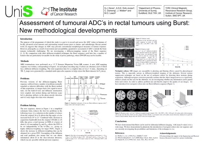
S 1Department of Physics, University of Surrey, Guildford, GU2 7XH, UK 2CRC Clinical Magnetic Resonance Research Group, Institute of Cancer Research, Sutton, SM2 5PT, UK S.J. Doran1, A.S.K. Dzik-Jurasz2, C. Domenig1, J. Wolber2 and M.O. Leach2 Assessment of tumoural ADC’s in rectal tumours using Burst: New methodological developments (a) (b) (d) (c) (a) (b) (c) Introduction The purpose of the programme of which this study is a part is to record and assess the ADC values in humans of locally advanced rectal tumours and surrounding normal tissues prior to chemo- and radiotherapy. Recent animal work [3] suggests that changes in ADC may precede conventional morphological measures of tumour response. However, principally as a result of movement and susceptibility, quantitative assessment of ADC’s outside the brain remains technically challenging. We are investigating a diffusion-mapping variant of the Burst sequence[1, 2]. By comparison with other diffusion-weighted techniques the Burst sequence used here has a number of features that are particularly favourable for the study of pelvic pathology, where DW-EPI may not be feasible. Figure 3: Volunteer study: Comparison of the old (left) and new (right) sequence in terms of SNR (matrix 128 128, FOV = 180 180 mm2). (e) Figure 4: Volunteer study: (a) - (c) 3 out of 7 diffusion-weighted images obtained with the new sequence in about 4 mins.; (d) calculated T2 map as it will be used for correcting the diffusion-weighted images for T2 related signal loss; (e) echo decay due to diffusion and T2, measured for three regions of interest (ROI) as indicated in (d) (matrix 128 128, FOV = 180 180 mm2). Methods MRI examinations were performed on a 1.5 T Siemens Magnetom Vision MR scanner. A new ADC-mapping sequence was written, corresponding to Figure1. At each phase-encoding step, 9 echoes are obtained, each of which has a different diffusion weighting. The total acquisition time for a complete data set was 1–4 mins., depending on TR. T2 maps were generated by a standard multi-spin-echo sequence to help correct the Burst datasets for T2 decay. Navigator echoes: MR images are susceptible to ghosting and blurring effects caused by physiological motion. This is especially serious in diffusion-weighted imaging of the abdomen. Several motion suppression techniques are based on the use of navigator echoes for detecting these motions during acquisition. Although navigator correction generally assumes that the object imaged moves as a rigid body, which is clearly not the case for the abdomen, we are investigating whether any information can be gained from such echoes. In order to address problem (iii) and thereby improve the quality of our images, we have introduced two navigator echoes into our Burst diffusion sequence (Figure 1). Problems Previous versions of the diffusion-mapping Burst sequence suffered from a number of problems normally regarded as inherent difficulties with the Burst method of data acquisition: (i) images had a low signal-to-noise ratio; (ii) the field-of-view and diffusion sensitisation were coupled; (iii) motion during and between scans led to ghosting artifacts in the phase-encode direction and (iv) only single-slice data were obtained. Figure 1: Simplified schematic of the new Burst-diffusion sequence with 7 image echoes and 2 navigator echoes. Problem Solving The new sequence, shown in Figure 1 in a simplified schematic form, reduces the first two problems in the following ways: (i) a reduction in the number of echoes (from the original 16 to 9) allows the flip angle a to be increased from 16 to 21. Combined with a decrease in acquisition bandwidth from the original 100 kHz to 25 kHz, this gives a total increase in SNR of a factor of about 4; (ii) uncoupling GR and Gdiff allows the FOV to be set independently of the b-value, i.e., giving us higher diffusion-weighting for larger FOVs. Figure 2 shows the increase in diffusion-weighting that can be obtained for a given FOV. Uncoupling of the diffusion and read gradients also means that we can obtain information about D in the slice-selection direction, which was not possible with the previous sequence.. Figure 5: Phantom study: (a) 6 out 7 diffusion-weighted images (matrix 128 128, FOV = 220 220 mm2), (b) surface plot of the navigator echo in the read direction, shown are the central 32 lines/columns, (c) navigator echo in the phase encode direction, displayed are again the central 32 lines/columns. Inserts show the 1D-FT of the raw navigator data. Conclusions We have demonstrated that Burst can be used for abdominal diffusion imaging, with many b-values over a wide range. We have demonstrated in phantoms the introduction of navigator echoes into our sequence and are currently investigating the possibilities and limitations of the technique in vivo. References [1] Wolber J., et al., [1999] Proc. ISMRM, 7, 1829 [2] Wheeler-Kingshott C. et al. [2000] Magn. Reson. Med., in press [3] Poptani H. et al. [1998] Canc. Gene. Ther., 5(2), 101-109 Acknowledgements CD acknowledges the support of the Engineering and Physical Sciences Research Council for a project studentship. Figure 2: The effect of uncoupling FOV and diffusion sensitisation: () Gread = Gdiff = 3.67 mT/m, b-values: 0.95 - 100 s/mm2; () Gread Gdiff, Gdiff = 10 mT/m, b-values: 0.11 - 1272 s/mm2(values for 128 128 matrix, FOV = 160 160 mm2).