Download
1 / 22
220 likes | 524 Views
Larynx Trachea & Bronchi. Prof. Saeed Abuel Makarem & Dr. Zeenat Zaidi. Objectives. At the end of the lecture, the students should be able to: Describe the Extent, structure and functions of the larynx. Describe the Extent, structure and functions of the Trachea.

E N D
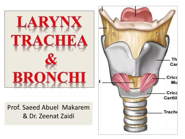
Larynx Trachea & Bronchi Prof. Saeed Abuel Makarem & Dr. Zeenat Zaidi
Objectives • At the end of the lecture, the students should be able to: • Describe the Extent, structure and functions of the larynx. • Describe the Extent, structure and functions of the Trachea. • Describe the bronchi and branching of the brochial tree. • Describe the functions of bronchi and their divisions.
Larynx • It is the part of respiratory tract containing the vocal cords. • Atube-shaped organ. • 2-inch-long • It opens into the laryngeal part of the pharynx above. • It is continuous below with thetrachea. • It is functioning in: • Deglutition (swallowing). • Respiration(breathing). • Phonation (phonation).
Relations • The larynx is related to major critical structures in the neck: • Carotid arteries , jugular veins, and vagus nerve. • Superior and inferior thyroid arteries. • Superior and recurrent laryngeal nerves.
Structure • The larynx consists of four basic components: • A cartilaginous skeleton. • Membranes and ligaments. • Muscles (intrinsic and extrinsic). • Mucosal lining.
Cartilages 3 3 1 • The cartilaginous skeleton is comprised of : • Thyroid. • Cricoid. Single • Epiglottis. • Arytenoid. • Corniculate. Paired • Cuneiform. • All the cartilages are hyaline , except the epiglottis. • Epiglottis is formed of elastic cartilage. • The cartilages are: • Connected by joints, ligaments & membranes. • Moved by muscles. 1 4 4 2 2 6 5
Membranes & Ligaments • Thyrohyoid membrane, (one median & two lateral thyrohyoid ligaments). • Median cricothyroid ligament. • Cricotracheal membrane. • Hyoepiglotticligament. • Thyroepiglottic ligament.
Quadrangular membrane: • Extends between the epiglottis and the arytenoid cartilages. • Its lower free margin forms the vestibular ligamentthat lies within the vestibular fold. • Cricothyroid membrane (conuselasticus): • Attached to upper border of the cricoid cartilage. • Its upper free margin forms vocal ligament.
Laryngeal Inlet • It is the upper opening of the larynx. • It is directed upward and backward and opens into the laryngeal part of the pharynx. • Bounded by: • Anteriorly: by the upper margin of epiglottis (E). • Posteriorly & below by arytenoid cartilages (A) • Laterally by aryepiglottic folds (AEF) • Piriform fossa: The area of the pharynx that surrounds the inlet of larynx. E AEF A
Laryngeal Cavity Rima vestibuli A • Extends from laryngeal inlet to the lower border of the cricoid cartilage. • Narrow in the region of the vestibular folds (rima vestibuli). • Narrowest in the region of the vocal folds (rimaglottidis). • Divided into three parts: • Supraglottic part, the part above the vestibular folds, is called the vestibule. • Ventricle: The part between the vestibular & the vocal folds. • Infraglottic part, the part below the vocal folds. B C Rima glottidis A B C
Muscles Divided into two groups: • Extrinsic muscles: divided into two groups • Elevators of the larynx • Depressors of the larynx • Intrinsic muscles: divided into two groups • Muscles controlling the laryngeal inlet • Muscles controlling the movements of the vocal cords Mucous Membrane • The cavity is lined with ciliated columnar epithelium • The surface of vocal folds, is covered with stratified squamous epithelium because of exposure to continuous trauma during phonation, • Contains many mucous glands, more numerous in the saccule (for lubrication of vocal folds)
Elevators of the Larynx • The Suprahyoid Muscles: • Digastric. • Stylohyoid. • Mylohyoid. • Geniohyoid. • The Longitudinal Muscles of the Pharynx: • Stylopharyngeus. • Salpingo-pharyngeus. • Palatopharyngeus. Depressors of Larynx • The Infrahyoid Muscles: • Sternohyoid. • Sternothyroid. • Omohyoid.
Muscles Controlling the Laryngeal Inlet • Oblique arytenoid • Aryepiglottic muscle
Muscle decreasing the Length & Tension of Vocal Cords Thyroarytenoid (vocalis) • Muscle increasing the Length & Tension of Vocal Cords • Cricothyroid: It is the only intrinsic muscle that present in the outer surface of the larynx.
Movements of the Vocal Cords • Adductors • Lateral cricoarytenoid • Transverse arytenoid • Abductor • Posterior cricoarytenoid Adduction Abduction
Nerve Supply • Sensory • Above the vocal cords: Internal laryngeal nerve, branch of the superior laryngeal of vagus. • Below the vocal cords: Recurrent laryngeal, of vagus. • Motor • All intrinsic muscles of the larynx are supplied by the recurrent laryngeal exceptcricothyroid, which is supplied by the theexternal laryngeal of superior laryngeal of vagus. Blood Supply • Arteries: • Upper half: Superior laryngeal artery, branch of superior thyroid artery. • Lower half: Inferior laryngeal artery, branch of inferior thyroid artery. • Veins: • Accompany the corresponding arterie.s • Lymphatics: • The lymph vessels drain into the deep cervical lymph nodes.
Trachea (windpipe) • Mobile, fibrocartilginous tube, 5 inches long, 1 inch in diameter. • Begins: In the neck below the cricoid cartilage of the larynx (at the level of C6). • Ends: below in the thorax at the level of sternal angle (lower border of T4), by dividing into right and left principal (main, primary) bronchi. • The ridge at the bifurcation is called carina.It is the most sensitive part of the tract and is associated with the cough reflex.
Relations in the Superior Mediastinum • Anterior • Sternum. • Thymus. • Left brachiocephalic vein. • Brachiocephalic artery. • Left common carotid a. • Arch of aorta. Left side • Arch of aorta. • Left common carotid artery. • Left subclavian a. • Left vagus & left phrenic nerves. • Pleura. Posterior • Esophagus. • Left recurrent laryngeal nerve. • Right side • Azygos vein. • Right vagus nerve. • Pleura.
Blood Supply • Arteries:Branches from the inferior thyroid andbronchial arteries. • Veins: Drain to inferior thyroid veins. Nerve Supply • Branches of vagusand therecurrent laryngeal nerves.(from vagus) supply sensory fibers to the mucous membrane. • Branches from the sympathetic trunks supply the trachealis muscle and the blood vessels. • Lymphatic Drainage • Into the pre- & paratracheallymph nodes.
Right Principal Bronchus About one inch long Wider, shorter and more vertical than the left ( foreign bodies). Gives superior lobar bronchus before entering the hilum of the right lung On entering the hilum it divides into middle and inferior lobar bronchi • Left Principal Bronchus • About two inches long • Narrower, longer and more horizontal than the right • Passes to the left below the arch of aorta and in front of esophagus • On entering the hilum of the left lung it divides into superior and inferior lobar bronchi
Bronchial Divisions • Within the lung each bronchus divides into number of branches that can be divided into two groups: Conduction zone branches • Primary (main) bronchi • Secondary (lobar) bronchi • Tertiary (segmental) bronchi (supply the bronchopulmonary segment) • Smaller bronchi • Bronchioles • Terminal bronchioles • Respiratory zone branches • Respiratory bronchioles • Alveolar ducts • Alveolar sacs • Alveoli
Thank You & Good Luck