Download
1 / 90
990 likes | 1.05k Views
Fundamentals of PACS. Harry H. Holdorf PhD, MPA, RDMS, RVT, LRT(AS). THE SKINNY PACS is the physical computer network DICOM is the set of rules that allows the components of the network to communicate. Contents. Definitions Introduction PACS DICOM Advantages of PACS

E N D
Fundamentals of PACS Harry H. Holdorf PhD, MPA, RDMS, RVT, LRT(AS)
THE SKINNY PACS is the physical computer network DICOM is the set of rules that allows the components of the network to communicate.
Contents Definitions Introduction PACS DICOM Advantages of PACS PACS fundamental parts Image acquisition Display workstations Archive Servers Workflow
Physician Review StationsTechnologist QC StationsFile Room/image Management StationsHanging ProtocolsImage Management FunctionsSummary/ConclusionPACS PROJECT
Definitions Digital Imaging and Communication in Medicine (DICOM) Teleradiology: The transmission of radiological patient images, such as x-rays, CTs MRIs, and Ultrasound Radiology Information System (RIS)
IntroductionWith the ever-increasing volume of ultrasound studies performed in a wide range of healthcare facilities, the ability to easily store and retrieve data helps radiologists and other physicians maintain an increased workflow while providing accurate diagnoses and quality of care for their patients. What can facilities do to make sure their ultrasound modalities and PACS communicate well, and what solutions are available to increase interoperability?
In the mid-1980s, early PACS development started, with limited availability of standards for exchanging imaging data between modalities and PACS. At the time, ultrasound units weren’t developed with any consideration for DICOM standards.
In 1994, the publication of the DICOM 3.0 standard resulted in a major improvement. Communication between PACS and modalities was now enabled by this standard, using Ethernet and the TCP/IP protocol. However, ultrasound modalities didn’t yet incorporate DICOM and digital output. It wasn’t until later that ultrasound manufacturers began to implement the DICOM standard in their products. Soon, improvements and new additions were added, first addressing 2D then 3D, 4D, and color data sets.
The ultrasound imaging data can be used to provide quantification of blood flow and anatomic measurements, which are an important aspect of the diagnostic process. As such, the ultrasound PACS should support this functionality. Often, hospitals will have a general PACS for all modalities but ultrasound and a PACS dedicated to ultrasound. Having interoperability between those two systems will assist the radiologist or specialist in viewing images from different imaging modalities like CT, MR, and DR, which again will improve productivity and quality of care.
Improving WorkflowPACS tries to achieve connected workflow throughout the medical care continuum, allowing physicians to improve workflow through many modalities. It can connect with ultrasound, cath diagnostic EKG [electrocardiogram], nuclear medicine, CT, MRI—all in one station—to improve access to the information. By connecting multiple modalities, it creates more efficiency.
An example of how interoperability can improve workflow is in having a Radiology Information System (RIS) that is interoperable with ultrasound. For example: If a medical center uses a RIS which is interfaced with both the PACS and the ultrasound machines. This improves workflow and accuracy because DICOM work-lists are generated through the RIS, which populates the ultrasound data fields automatically without a technologist having to retype information.
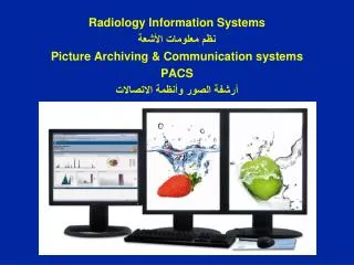
Picture Archiving and Communication Systems (PACS) As imaging departments move from film-based acquisition and archiving (hard-copy film and document storage) to digital acquisition and archiving (soft-copy storage), a complex computer network has been created to manage images. This network is called Picture Archiving and Communication Systems (PACS) and can be likened to a “virtual film library.” Images stored on digital media are housed in PACS archives.
PACS is a sophisticated array of hardware and software that can connect all modalities with digital output (nuclear medicine, ultrasound, computed tomography, magnetic resonance imaging, angiography, mammography, and radiography). The acronym PACS can best be explained as follows: P—Picture: the digital medical image(s) A—Archiving: the “electronic” storage of the images C—Communication: the routing (retrieval/sending) and displaying of the images S—System: the specialized computer network that manages the complete system
A PACS can accept any image that is in digital imaging and communications in medicine (DICOM) format, for which it is set up to receive, whether it is from cardiology, radiology, or pathology. A PACS serves as the file room, reading room, duplicator, and courier. It can provide image access to multiple users at the same time, on-demand images, electronic annotation of images, and specialty image processing.
A PACS is often custom designed for a facility. The software is generally the same, but the components are arranged differently. Specific factors are involved in designing a PACS for an institution, such as the volume of patients, the number of areas where images are interpreted, the locations where images are viewed by physicians other than radiologists, and the money available for purchase.
The connection of various equipment types and modalities to a PACS is complex. Standards have been developed to ensure that all manufacturers and types of equipment are able to communicate and effectively transmit images and information. Current standards include DICOM (Digital Imaging Communications in Medicine) and HL7 (Health Care Level 7). Although standards may not always provide for an instantaneous functionality between devices, they do allow for resolution of connectivity problems.
DICOM is universally accepted industry standard for transferring radiologic images and their medical information between computers.
The HL-7 standard oversees most clinical and administrative data such as demographics, reports, claims, and orders. As with DICOM, HL-7 is composed of many parts and is used at many levels within various hospital systems. It is the standard generally used in communication between the hospital information system (HIS) and the radiology information system (RIS). The HIS holds the patient’s full medical information, from hospital billing to the inpatient ordering system. The RIS holds all radiology-specific patient data, from the patient scheduling information to the radiologist’s dictated and transcribed report.
Digital Imaging and Communications in Medicine (DICOM) is a communication standard for information sharing between PACS and imaging modalities. Health Level Seven standard (HL7) is a communication standard for medical information.
For optimum efficiency, the PACS should be integrated with the Radiology Information System (RIS) or the Hospital Information System (HIS). Because these information systems support the operations of an imaging department through exam scheduling, patient registration, report archiving, and film tracking, integration with PACS maintains integrity of patient data and records and promotes overall efficiency.
When a PACS is used, instead of hard-copy radiographs that must be processed, handled, viewed, transported, and stored, the soft-copy digital images are processed with the use of a computer, viewed on a monitor, and stored electronically. Most PACS use web browsers to enable easy access to the images by users from any location. Physicians may view these radiologic images from a personal computer at virtually any location, including their home.
ADVANTAGES OF PACS: • Elimination of less efficient traditional film libraries and their inherent problem of physical space requirements for hard-copy images • Convenient search for and retrieval of images • Rapid (electronic) transfer of images within the hospital (e.g., clinics, operating rooms, treatment units) • Ease in consulting outside specialists—teleradiology. Teleradiography is the electronic transmission of diagnostic images from one location to another for purposes of interpretation and/or consultation. • Simultaneous viewing of images at multiple locations • Elimination of misplaced, damaged, or missing films • Increase in efficiency of reporting exams with soft-copy images (compared with hard-copy images) • Reduction of the health and environmental impact associated with chemical processing, as a result of decreased use
In the mid to early 1980s, different versions of PACS were being developed, primarily by research and academic institutions. They were homegrown and usually involved one or possibly two modalities. These early systems were hard to put together because there was little standardization in image formats. Each vendor had its own proprietary way of archiving images, and there was little need or desire to share archiving methods. Once DICOM (standards that allow imaging modalities and PACS to communicate in the same “language”) was established, more vendors began using it to communicate between modalities and PACS. Full-scale acceptance of DICOM was pushed by the consumer to make it possible for equipment from different manufacturers to talk to each other. The first full-scale PACS in the United States was installed at the VA Medical Center in Baltimore in 1993. Their PACS covered all modalities except mammography. Soon after installing their PACS, the Baltimore Medical Center asked the vendor to interface to their radiology information system (RIS), hospital information system (HIS), and electronic medical record (EMR).
PACS fundamental parts: • Image acquisition • Display workstations • Archive servers
Image Acquisition In modern radiology departments, most images are acquired in a digital format, meaning that the images are inherently digital and can be transferred via a computer network. Ultrasound, computed tomography (CT), magnetic resonance imaging (MRI), and nuclear medicine have been digital for many years and have been taking advantage of PACS far longer than general radiography has
Display Workstations A display workstation is any computer that a health care worker uses to view a digital image. It is the most interactive part of a PACS, and these workstations are used inside and outside of radiology. The display station receives images from the archive or from the various radiology modalities and presents them for viewing. The display workstation has PACS application software that allows the user to perform minor image-manipulation techniques to optimize the image being viewed. Some display stations have advanced software to perform more complex image-manipulation techniques
The display workstation is the most interactive part of a PACS, consisting of a monitor and a computer with a mouse and keyboard. In addition, each system has hardware that fits the users’ requirements. As you know, conventional film/screen radiography uses large multiviewer lightboxes to display the images . Early in the history of PACS, radiologists believed that they needed four to six monitors to match the viewing capability they had with the lightboxes. As the radiologists have become more comfortable viewing images on monitors, the number of monitors required by the radiologists has decreased to an average of two
The monitor is one of the most important elements of a PACS display station. The cathode ray tube (CRT) and the liquid crystal display (LCD) are the most popular types of monitors in a radiology department. The LCD has decreased in price and increased in quality during the past few years and will soon take over the entire PACS display market because of its size, resolution, and lack of heat production. The LCD also requires less maintenance, gives out more light, and can be used in areas with a high amount of ambient light. In early PACS reading rooms, supplemental air conditioning had to be installed to offset the heat put out by multiple CRTs. Along with the number of monitors used, the resolution and orientation of the monitor are also factors in determining which type of monitor to buy for each workstation.