Download
1 / 24
250 likes | 313 Views
Development of Respiratory System. BY : Dr.SANA ALSHAARAWY. OBJECTIVES. At the end of the lecture the students should be able to: Identify the development of the laryngeotracheal (respiratory) diverticulum . Identify the development of the larynx. Identify the development of the trachea.

E N D
Development of Respiratory System BY : Dr.SANA ALSHAARAWY
OBJECTIVES At the end of the lecture the students should be able to: Identify the development of the laryngeotracheal (respiratory) diverticulum. Identify the development of the larynx. Identify the development of the trachea. Identify the development of the bronchi & Lungs. Describe the periods of the maturation of the lung. Identify the most congenital anomaly.
Respiratory System • Upper respiratory tract: • Nose • Nasal cavity & paranasal sinuses • Laryngo-pharynx • Larynx. • Lower respiratory tract: • Trachea • Bronchi • Lungs
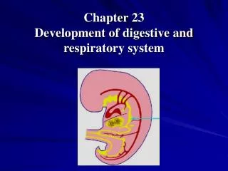
Development of the Respiratory Tract • Begins during the 4th week of development. • Begins as a median outgrowth (laryngo-tracheal groove) from the caudal part of the ventral wall of the primitive pharynx (foregut) • The groove envaginates and forms the laryngotracheal (respiratory) diverticulum.
A longitudinal tracheo-esophageal septum develops and divides the diverticuluminto a: • Dorsal portion: primordium of the oropharynx and esophagus • Ventral portion: primordium of larynx, trachea, bronchi and lungs
The proximal part of the respiratory diverticulum remains tubularand forms larynx & trachea. The distal end of the diverticulum dilates to form lung bud, which divides to give rise to 2lung buds (primary bronchial buds)
Theendodermlining thelaryngotrachealdiverticulum (Respiratory diverticulum) gives rise to the: Epithelium & Glandsof the respiratory tract. The surroundingsplanchnic mesodermgives rise to the: Connective tissue, Cartilage & Smooth muscles of the respiratory tract
Development of the Larynx • The opening of the laryngotrachealdiverticuluminto the primitive foregut becomes the laryngeal orifice. • The epithelium & glands are derived from endoderm. • Laryngeal muscles & the cartilages of the larynx exceptEpiglottis develop from the mesoderm of 4th& 6th pairs of pharyngeal arches.
Epiglottis • It develops from the caudal part of the hypopharyngeal eminence, a swelling formed by the proliferation of mesoderm in the floor of the pharynx. Growth of the larynx and epiglottis is rapid duringthe first three years after birth. By this time the epiglottis has reached its adult form.
Recanalization of larynx The laryngeal epithelium proliferates rapidly resulting in temporary occlusion of the laryngeal lumen Recanalization of larynx normally occurs by the 10th week. Laryngeal ventricles, vocal folds and vestibular folds are formed during recanalization.
Development of the Trachea • The endodermal lining of the laryngotracheal tube distal to the larynx differentiates into the epithelium and glands of the trachea and pulmonary epithelium • The cartilages, connective tissue, and muscles of the trachea are derived from the mesoderm.
Development of the Bronchi & Lungs • The 2 primary bronchial buds grow laterally into the pericardio-peritoneal canals (part of intra-embryonic celome), which the primordia of pleural cavities • Bronchial buds divide and re-divide to give the bronchial tree.
The right main bronchus is slightly larger (wider) than the left one and is oriented more vertically • The embryonic relationship persists in the adult. • The main bronchi subdivide into secondary and tertiary (segmental)bronchi which give rise to further branches.
The segmental bronchi10 in right lung and 8 or 9 in the left lung begin to form by the 7th week • The surrounding mesenchyme also divides. • Each segmental bronchus with its surrounding mass of mesenchymeis the primordium of a bronchopulmonary segment.
Development of the pleura • As the lungs develop they acquire a layer of visceral pleura from splanchnic mesenchyme. • The thoracic body wall becomes lined by a layer of parietal pleura derived from the somatic mesoderm.
Maturation of the Lungs • Maturation of lung is divided into 4 periods: • Pseudoglandular (5 - 17 weeks) • Canalicular (16 - 25 weeks) • Terminal sac (24 weeks - birth) • Alveolar (late fetal period - childhood) • These periods overlap each other because the cranial segments of the lungs mature faster than the caudal ones (16 - 25 weeks) (24 weeks - birth) (late fetal period - childhood) (5 - 17 weeks)
Pseudoglandular Period (5-17 weeks) • Developing lungs somewhatresemblesan exocrine gland during this period. • By 17 weeks all major elements of the lung have formed except those involved with gas exchange (alveoli). • Respiration is NOT possible. • Fetusesborn during this period are unable to survive.
Canalicular Period (16-25 weeks) • Lung tissue becomes highly vascular. • Lumina of bronchi and terminal bronchioles become larger. • By 24 weeks each terminal bronchiole has given rise to two or more respiratory bronchioles. • The respiratory bronchioles divide into 3 to 6 tubular passages called alveolar ducts. • Somethin-walled terminal sacs (primordial alveoli) developat the end of respiratory bronchioles. • Respiration is possible at the end of this period. • Fetus born at the end of this period may survive if given intensive care (but usually die because of the immaturity of respiratory as well as other systems)
Terminal Sac Period (24 weeks - birth) • Many more terminal sacs develop. • Their epithelium becomes very thin. • Capillaries begin to bulge into developing alveoli. • The epithelial cells of the alveoli and the endothelial cells of the capillaries come in intimate contact and establish the blood-air barrier. • Adequate gas exchange can occur which allows the prematurely born fetus to survive
By 24 weeks, the terminal sacs are lined by: • Squamoustype I pneumocytesand • Rounded secretory, type II pneumocytes, that secrete a mixture of phospholipids called surfactant. Surfactant production begins by 20 weeks and increases during the terminal stages of pregnancy. Sufficient terminal sacs, pulmonary vasculature & surfactant are present to permit survival of a prematurely born infants Fetuses born prematurely at 24-26 weeks may suffer from respiratory distress due to surfactant deficiencybutmay surviveif given intensive care.
Alveolar Period (32 weeks – 8 years) • At the beginning of the alveolar period, each respiratory bronchioleterminates in a cluster of thin-walled terminal saccules separated from one another by loose connective tissue. • These terminal sacculesrepresent future alveolar sacs.
Characteristic mature alveoli do not form until after birth, so; 95% of alveoli develop postnatally. • About 50 million alveoli, one sixth of the adult number are present in the lungs of a full-term newborn infant. • From 3-8 year or so, the number of alveoli continues to increase, forming additional primordial alveoli. • By about the eighth year, the adult complement of 300 million alveoli is present.
Developmental anomaliesTracheo-esophageal Fistula • An abnormal passage between the trachea and esophagus. • Results fromincomplete division of the cranial part of the foregut into respiratory and esophageal parts by the tracheo-esophageal septum. • Occurs once in 3000 to 4500 live births. • Most affected infants are males. • In more than 85% of cases, the fistula is associated with esophageal atresia(esophagus ends in a blind-ended pouch rather than connecting normally to the stomach).