1 / 36

Figure 43.0 Specialized lymphocytes attacking a cancer cell

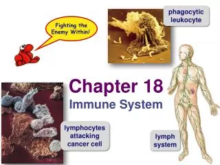
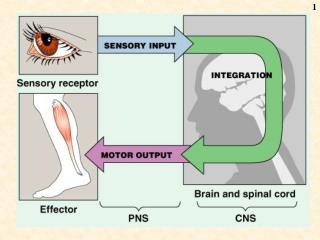
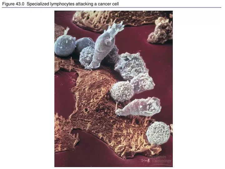
Figure 43.0 Specialized lymphocytes attacking a cancer cell. Figure 43.1 An overview of the body's defenses. Figure 43.2 First-line respiratory defenses. Figure 43.3 Phagocytosis by a macrophage. Figure 43.3x Macrophage. Figure 43.x1 Anabaena phagocytosed by a human neutrophil.

rredmon
Download Presentation

Figure 43.0 Specialized lymphocytes attacking a cancer cell

An Image/Link below is provided (as is) to download presentation Download Policy: Content on the Website is provided to you AS IS for your information and personal use and may not be sold / licensed / shared on other websites without getting consent from its author. Content is provided to you AS IS for your information and personal use only. Download presentation by click this link. While downloading, if for some reason you are not able to download a presentation, the publisher may have deleted the file from their server. During download, if you can't get a presentation, the file might be deleted by the publisher.

E N D

Presentation Transcript


  1. Figure 43.0 Specialized lymphocytes attacking a cancer cell

  2. Figure 43.1 An overview of the body's defenses

  3. Figure 43.2 First-line respiratory defenses

  4. Figure 43.3 Phagocytosis by a macrophage

  5. Figure 43.3x Macrophage

  6. Figure 43.x1 Anabaena phagocytosed by a human neutrophil

  7. Figure 43.4 The human lymphatic system

  8. Figure 43.5 A simplified view of the inflammatory response

  9. Figure 43.6 Clonal selection

  10. Figure 43.7 Immunological memory

  11. Figure 43.8 The development of lymphocytes

  12. Figure 43.8x B lymphocyte

  13. Figure 43.9 The interaction of T cells with MHC molecules

  14. Figure 43.10 An overview of the immune responses (Layer 1)

  15. Figure 43.10 An overview of the immune responses (Layer 2)

  16. Figure 43.10 An overview of the immune responses (Layer 3)

  17. Figure 43.10 An overview of the immune responses (Layer 4)

  18. Figure 43.11 The central role of helper T cells: a closer look

  19. Figure 43.12a The functioning of cytotoxic T cells

  20. Figure 43.12b A cytotoxic T cell has lysed a cancer cell

  21. Figure 43.13 Humoral response to a T-dependent antigen (Layer 1)

  22. Figure 43.13 Humoral response to a T-dependent antigen (Layer 2)

  23. Figure 43.13 Humoral response to a T-dependent antigen (Layer 3)

  24. Figure 43.14 Epitopes (antigenic determinants)

  25. Figure 43.15a,b The structure of a typical antibody molecule

  26. Figure 43.15c Antibody molecule

  27. Table 43.1 The Five Classes of Immunoglobulins

  28. Figure 43.16 Effector mechanisms of humoral immunity

  29. Figure 43.17 The classical complement pathway, resulting in lysis of a target cell

  30. Figure 43.18 Mast cells, IgE, and the allergic response

  31. Figure 43.x4 Alternaria spores, a cause of allergies in humans

  32. Figure 43.19 A T cell infected with HIV

  33. Figure 43.19x1 HIV on a lymphocyte, detail

  34. Figure 43.19x2 HIV budding

  35. Figure 43.19x2a T cell infected with HIV

  36. Figure 43.20 The stages of HIV infection

More Related