Download
1 / 23
230 likes | 255 Views
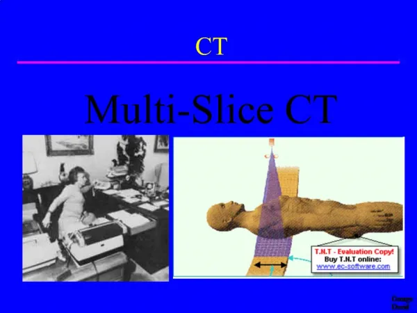
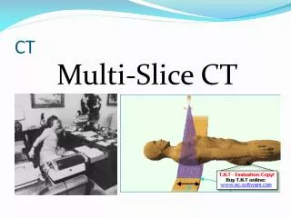
Single Slice Spiral - Helical CT. Oh no, not more physics…. Spiral CT. Incentives for development Shorter study times Improved 3D imaging New technology required Slip ring Allows continuous gantry rotation. Conventional (Non-spiral) CT. Tube rotates once around patient Table stationary

E N D
Single Slice Spiral - Helical CT Oh no, not more physics…
Spiral CT • Incentives for development • Shorter study times • Improved 3D imaging • New technology required • Slip ring • Allows continuous gantry rotation
Conventional (Non-spiral) CT • Tube rotates once around patient • Table stationary • data for one slice collected • Table increments one slice thickness • Repeat • Tube rotates opposite direction
Volume Scanning • Data collected continuously • Table moves continuously • Focal spot traces spiral pathwith respect to patient
Helical Reconstruction Complication • Patient moves as gantry rotates • No two fan beams at same z coordinate “z” direction
As Gantry Rotates,Fan Angles Repeat • Distance between repetitions is movement of table during one rotation “z” direction
Data Acquisition Challenges • Projection data not confined to single slice • Streak artifacts • caused by motion • special algorithms required Position at start of rotation Position at start of rotation Position of interest
Calculating Fan Beams at Odd Locationsusing Interpolation • No complete data set for any single z location • Use 2 closest beams in correct orientation • Calculate beam attenuation by interpolating between adjacent beams “z” direction
Spiral Reconstruction Algorithms = real data point • Uses interpolation for • input projection data • output slice attenuation data • Slice can be calculated at any position from raw projection data coordinate of interest Interpolated data
Disadvantage of Interpolation Trust me, interpolation is a guess • Can increase effective slice thickness • Calculation averages data measured at many z values “z” direction
Data Acquisition Challenges • No single slice defined by acquisition geometry • slice localization more difficult • Different slice volume geometry • conventional: cylinder • spiral: wafer with radial crack • Slight increase in effective slice thickness • slice thickness influenced by • fan beam thickness • speed of table motion
Table Moves During Helical Scanning table increment during one rotation Slice Pitch = --------------------------------------- slice thickness Slice thickness TableIncrement
Table Moves During Helical Scanning • Slice thickness determined by collimation • Table motion per revolution determined by table speed table motion during one rotation Slice Pitch = --------------------------------------- slice thickness Slice thickness TableIncrement
Single-Slice Detectors • Many detectors rotate around patient • Single row in z-direction • Slice thickness determined by collimation SliceThickness Z-Axis
Pitch = 1 • Pitch = 1 means patient moves exactly one slice thickness per revolution of tube table motion during one tube rotation Slice Pitch = ---------------------------------------------- slice thickness Beam positions when tube directly above patient
Pitch <1 • Pitch < 1 means patient moves less than a slice thickness during one tube rotation • Can improve visualization of objects table motion during one tube rotation Slice Pitch = ----------------------------------------------- slice thickness Beam positions when tube directly above patient
Pitch >1 • Pitch > 1 means patient moves further than slice thickness during one tube rotation table motion during one tube rotation Slice Pitch = ---------------------------------------------- slice thickness Beam positions when tube directly above patient
Pitch >1 • No gap in image coverage when viewing study table motion during one tube rotation Slice Pitch = ---------------------------------------------- slice thickness No Gap Beam position when tube directly belowpatient
Spiral vs. Conventional CT & Patient Dose • Dose is strongly dependent on pitch Please explain. Inquiring minds wanna know
Pitch = 1 • equivalent dose to non-spiral
Pitch >1 • lower dose for spiral • Table moves faster • Table increment per tube rotation > one slice thickness
Pitch <1 • higher dose for spiral • Table moves slower • table increment per tube rotation < one slice thickness
Pitch & Dose • Dose inversely proportional to pitch • Pitch = 0.5 => Dose doubles • Pitch = 2 => Dose cut in half