Download
1 / 47
470 likes | 619 Views
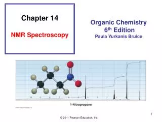
13. Structure Determination: Nuclear Magnetic Resonance Spectroscopy. Based on McMurry’s Organic Chemistry , 7 th edition. The Use of NMR Spectroscopy. Used to determine relative location of atoms within a molecule Most helpful spectroscopic technique in organic chemistry

E N D
13. Structure Determination: Nuclear Magnetic Resonance Spectroscopy Based on McMurry’s Organic Chemistry, 7th edition
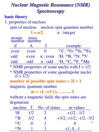
The Use of NMR Spectroscopy • Used to determine relative location of atoms within a molecule • Most helpful spectroscopic technique in organic chemistry • Maps carbon-hydrogen framework of molecules • Depends on very strong magnetic fields (imagine the strongest electromagnet you can and the imagine it on steroids)
Why This Chapter? • NMR is the most valuable spectroscopic technique used for structure determination • More advanced NMR techniques are used in biological chemistry to study protein structure and folding
13.1 Nuclear Magnetic Resonance Spectroscopy • 1H or 13C nucleus spins and the internal magnetic field aligns parallel to or against an aligned external magnetic field (See Figure 13.1) • Parallel orientation is lower in energy making this spin state more populated • Radio energy of exactly correct frequency (resonance) causes nuclei to flip into anti-parallel state • Energy needed is related to molecular environment (proportional to field strength, B) – see Figure 13.2
The spin state of a nucleus is affected by an applied magnetic field
The energy difference between the two spin states depends on the strength of the magnetic field (that the atom “feels”)
absorb DE a-spin states b-spin states release DE Signals detected by NMR FID
The electrons surrounding a nucleus affect the effective magnetic field sensed by the nucleus Electron poor environment Electron rich environment
Shielded nuclei do not ‘sense’ as large a magnetic field as deshielded nuclei do. As a result, the energy difference between the - and -spin states is much lower in energy for shielded nuclei and resonate at a lower frequency. Deshielded nuclei have a much higher energy difference between the - and -spin states and these resonate at a much higher frequency.
13.2 The Nature of NMR Absorptions • Electrons in bonds shield nuclei from magnetic field • Different signals appear for nuclei in different environments
The NMR Measurement • The sample is dissolved in a solvent that does not have a signal itself* and placed in a long thin tube • The tube is placed within the gap of a magnet and spun • Radiofrequency energy is transmitted and absorption is detected • Species that interconvert give an averaged signal that can be analyzed to find the rate of conversion • Can be used to measure rates and activation energies of very fast processes
13.3 Chemical Shifts • The relative energy of resonance of a particular nucleus resulting from its local environment is called chemical shift • NMR spectra show applied field strength increasing from left to right • Left part is downfield the right is upfield • Nuclei that absorb on upfield side are strongly shielded • Chart calibrated versus a reference point, set as 0, tetramethylsilane [TMS]
Measuring Chemical Shift • Numeric value of chemical shift: difference between strength of magnetic field at which the observed nucleus resonates and field strength for resonance of a reference • Difference is very small but can be accurately measured • Taken as a ratio to the total field and multiplied by 106 so the shift is in parts per million (ppm) • Absorptions normally occur downfield of TMS, to the left on the chart • Calibrated on relative scale in delta () scale • Independent of instrument’s field strength
13.4 13C NMR Spectroscopy: Signal Averaging and FT-NMR • Carbon-13: only carbon isotope with a nuclear spin • Natural abundance 1.1% of C’s in molecules • Sample is thus very dilute in this isotope • Sample is measured using repeated accumulation of data and averaging of signals, incorporating pulse and the operation of Fourier transform (FT-NMR) • All signals are obtained simultaneously using a broad pulse of energy and resonance recorded • Frequent repeated pulses give many sets of data that are averaged to eliminate noise • Fourier-transform of averaged pulsed data gives spectrum (see Figure 13-6)
1 scan of conc. sample 200 scans of same sample Fig. 13-6, p. 447
13.5 Characteristics of 13C NMR Spectroscopy • Provides a count of the different types of environments of carbon atoms in a molecule • 13C resonances are 0 to 220 ppm downfield from TMS (Figure 13-7) • Chemical shift affected by electronegativity of nearby atoms • O, N, halogen decrease electron density and shielding (“deshield”), moving signal downfield. • sp3 C signal is at 0 to 9; sp2 C: 110 to 220 • C(=O) at low field, 160 to 220
13C NMR Low Field High Field Deshielding Shielding Down field Up field 1H NMR
Spectrum of 2-butanone is illustrative- signal for C=O carbons on left edge
13.6 DEPT 13C NMR Spectroscopy • Improved pulsing and computational methods give additional information • DEPT-NMR (distortionless enhancement by polarization transfer) • Normal spectrum shows all C’s then: • Obtain spectrum of all C’s except quaternary (broad band decoupled) • Change pulses to obtain separate information for CH2, CH • Subtraction reveals each type (See Figure 13-10)
DEPT 13C NMR distinguish among CH3, CH2, and CH Groups (Distortionless Enhancement by Polarization Transfer
13.7 Uses of13C NMR Spectroscopy • Provides details of structure • Example: product orientation in elimination from 1-chloro-methyl cyclohexane • Difference in symmetry of products is directly observed in the spectrum • 1-chloro-methylcyclohexane has five sp3 resonances ( 20-50) and two sp2resonances 100-150
13.8 1H NMR Spectroscopy and Proton Equivalence • Proton NMR is much more sensitive than 13C and the active nucleus (1H) is nearly 100 % of the natural abundance • Shows how many kinds of nonequivalent hydrogens are in a compound • Theoretical equivalence can be predicted by seeing if replacing each H with “X” gives the same or different outcome • Equivalent H’s have the same signal while nonequivalent are “different” and as such may cause additional splitting (diastereotopic effect) • There are degrees of nonequivalence
Nonequivalent H’s • Replacement of each H with “X” gives a different constitutional isomer • Then the H’s are in constitutionally heterotopic environments and will have different chemical shifts – they are nonequivalent under all circumstances
Equivalent H’s • Two H’s that are in identical environments (homotopic) have the same NMR signal • Test by replacing each with X • if they give the identical result, they are equivalent • Protons are considered homotopic
Enantiotopic Distinctions • If H’s are in environments that are mirror images of each other, they are enantiotopic • Replacement of each H with X produces a set of enantiomers • The H’s have the same NMR signal (in the absence of chiral materials)
Diastereotopic Distinctions • In a chiral molecule, paired hydrogens can have different environments and different shifts • Replacement of a pro-R hydrogen with X gives a different diastereomer than replacement of the pro-S hydrogen • Diastereotopic hydrogens are distinct chemically and spectrocopically
13.9 Chemical Shifts in 1H NMR Spectroscopy • Proton signals range from 0 to 10 • Lower field signals are H’s attached to sp2C • Higher field signals are H’s attached to sp3C • Electronegative atoms attached to adjacent C cause downfield shift
13.10 Integration of 1H NMR Absorptions: Proton Counting • The relative intensity of a signal (integrated area) is proportional to the number of protons causing the signal • This information is used to deduce the structure • For example in ethanol (CH3CH2OH), the signals have the integrated ratio 3:2:1 • For narrow peaks, the heights are the same as the areas and can be measured with a ruler
13.11 Spin-Spin Splitting in 1H NMR Spectra • Peaks are often split into multiple peaks due to interactions between nonequivalent protons on adjacent carbons, called spin-spin splitting • The splitting is into one more peak than the number of H’s on the adjacent carbon (“n+1 rule”) • The relative intensities are in proportion of a binomial distribution and are due to interactions between nuclear spins that can have two possible alignments with respect to the magnetic field • The set of peaks is a multiplet (2 = doublet, 3 = triplet, 4 = quartet)
Simple Spin-Spin Splitting • An adjacent CH3 group can have four different spin alignments as 1:3:3:1 • This gives peaks in ratio of the adjacent H signal • An adjacent CH2 gives a ratio of 1:2:1 • The separation of peaks in a multiplet is measured is a constant, in Hz • J (coupling constant)
Rules for Spin-Spin Splitting • Equivalent protons do not split each other • The signal of a proton with n equivalent neighboring H’s is split into n + 1 peaks • Protons that are farther than two carbon atoms apart do not split each other
10.0 9.0 8.0 7.0 6.0 5.0 4.0 3.0 2.0 1.0 0 BrCH2CH3 4 lines; quartet 3 lines; triplet CH3 CH2 Chemical shift (, ppm)
10.0 9.0 8.0 7.0 6.0 5.0 4.0 3.0 2.0 1.0 0 BrCH(CH3)2 2 lines; doublet 7 lines; septet CH3 CH Chemical shift (, ppm)
Protons Bonded to Oxygen and Nitrogen These protons can undergo proton exchange The greater the extent of the hydrogen bond, the greater the chemical shift They always appear as broad signals
dry ethanol ethanol with acid
To observe well-defined splitting patterns, the difference in the chemical shifts (in Hz) must be 10 times the coupling constant values
1H NMR Spectra of 2-sec-butylphenol at Different Field Strengths 60 MHz 300 MHz
13.12 More Complex Spin-Spin Splitting Patterns • Spectra can be more complex due to overlapping signals, multiple nonequivalence • Example: trans-cinnamaldehyde
13.13 Uses of 1H NMR Spectroscopy • The technique is used to identify likely products in the laboratory quickly and easily • Example: regiochemistry of hydroboration/oxidation of methylenecyclohexane • Only that for cyclohexylmethanol is observed
Peaks in a 13C NMR spectrum are typicallysinglets 13C—13C splitting is not seen because the probability of two 13C nuclei being in the same molecule is very small. 13C—1H splitting is not normally seen because spectrum is measured under conditions that suppress this splitting (broadband decoupling).
1H Decoupled and Coupled 13C Spectra of 2-butanol decoupled coupled