Download
1 / 35
490 likes | 1.24k Views
Pediatric Metabolic Bone Disease. Bryce Nelson, MD/PhD Pediatric Endocrinology Greenville Hospital System SEACSM Meeting, Clinical Track Program 2/10/12. Objectives. Discuss contributors to pediatric bone disease Discuss evaluation of child with fragility fractures

E N D
Pediatric Metabolic Bone Disease • Bryce Nelson, MD/PhD • Pediatric Endocrinology • Greenville Hospital System • SEACSM Meeting, Clinical Track Program • 2/10/12
Objectives • Discuss contributors to pediatric bone disease • Discuss evaluation of child with fragility fractures • Discuss treatment options for children with bone disease
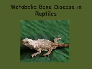
Bone Health in Children • Osteoporosis in adults considered a pediatric disease (Dent, et. al. Postgrad Med J. 1973) • Bone Mass achieved in adolescence is main contributor of peak bone mass which is major determinant of fracture risk
Fragility vs. Traumatic Fracture • Vertebral fractures and femur fractures without significant trauma • Infant fractures? Abuse or not? • Immobilization
Fractures: Tansient Fragility? • Fracture incidence proportional to height velocity • Age 11-12 in girls • Age 13-14 in boys • Peak bone mass lags behind peak growth velocity by about 18 months
Peak Bone Mass • Bone Mineral Density >95% of peak value by age 20 • First at hip, then spine, then whole body • Gender Difference • Earlier in women then men
Risk Factors for Low Bone Mineral Density • Genetics (60-80%) • Physical Activity (10-20%) • Environmental (calcium, vitamin D intake, drug induced)
Some Disorders Associated with Fragility Fractures • Primary Conditions • Genetic Disorders • Osteogenesis Imperfecta • Idiopathic Juvenile Osteoporosis • Chronic Inflammatory • SLE • Inflammatory Bowel Disease • Immobilization • Infiltrative • Leukemia • Endocrine • Hypogonadism, GH deficiency, Cushing, Hyperthyroidism, Diabetes • Nutritional • Vitamin D Deficiency, celiac disease, cystic fibrosis, anorexia • Renal • Chronic Kidney Disease • Iatrogenic • Glucocorticoids, anticonvulsants, methotrexate, radiation, antiretroviral
To make the issue more complicated… • Children >8 years of age do not achieve RDI of Ca • Adequate intake affected by age, gender, physical activity and diet • Calcium RDI varies with age Greer, FR et. al Pediatrics. 117. 2006. 578-585
Vitamin D Metabolism • 7-dehydrocholesterol converted to Vitamin D3 by UV • Converted to 25-OH-VitD3 in liver • Active form 1,25OH-Vitamin D3 in kidney • 1-alpha-hydroxylase • PTH • Circulates in blood bound to either DBP or albumin • Little free form in blood http://www.mja.com.au
Vitamin D: Is it our new snake oil?…more than just rickets • Vitamin D deficiency or insufficiency often seen in post-menopausal women and older Americans with osteoporosis • May be protective against some cancers • Asthma • Multiple Sclerosis • Crohn’s Disease • Ulcerative Colitis
Risk Factors for Vitamin D Deficient Rickets • Poor sunlight exposure • Poor dietary intake of Vitamin D • No vitamin supplementation • Breast fed infants, particularly non-Caucasians • Females • Low Socioeconomic Status • Low BMI or high BMI • Elderly • African American, Hispanic, or Middle Eastern descent • Chronic illness, malabsorption, renal or liver disease • Living during the winter!
Vitamin D Levels Wagner, CL, et al. Pediatrics. 2008. 1142.
History & Physical • Breast fed • Race • Metaphyseal cupping and fraying • Genu valgum or varum • Rachitic rosary • Frontal bossing
Lab evaluation • Second Tier Labs • Bone Turnover Markers • Osteocalcin • Urine N-telo peptides • Bone Marrow • First Tier Labs • CBC, diff, platelets • CMP (alkaline phosphatase) • Sed rate • PTH • Ca, Mg, PO4 • Spot urine Ca/Cr ratio • 25 OH vitamin D
Bone Densitometry in Children • Quantitative CT (volumetric) • Dual energy X-ray Absorptiometry (DXA, areal density)
DXA in Children • Advantages: fast, low radiation exposure, reasonable image resolution • Disadvantages: body composition changes, limited reference data, puberty, stature effects
Areal vs Volumetric BMD DXA underestimates total areal BMD in short children or overestimates in tall or “big bone” courses.washington.edu/bonephys/opBMAD.html
WHO Classification of Bone Mineral Density (BMD) • No densitometric criteria in children for osteoporosis • Z score -2.0 or less: “low BMD for age” • Z score needs to be bone age and stature adjusted • Spine and total body are preferred skeletal sites for measurement
Consideration and Controversy • Osteoporosis diagnosis in children requires both clinically significant fracture history and low BMD • No link between vitamin D and fracture risk in children • DXA needs to be performed appropriately
Basic Treatment • Identify and treat any underlying cause • Maximize calcium and vitamin D or replete if deficient • Weight bearing physical activity when appropriate
US Recommended Daily Ca intake Institute of Medicine, Food and Nutrition Board, Dietary References for Intakes for Calcium, Phosphorus, Magnesium, Vitamin D, and Fluoride. National Academy Press. 1997
AAP Recommendations • ALL breastfed infants and formula fed infants taking <1L/day should take 400 IU vit D supp, to be started within first few days of life • Children and adolescents without appropriate sun exposure AND less than 500 ml of vit D-milk per day should also take vit D supp (400 IU/d) • Premature infants to be started on 400-800 IU/day at birth Misra, M et. al Pediatrics. 122. 2008. 398-417
Endocrine Society GuidelinesVitamin D Deficiency Replacement * Patients on anticonvulsants, glucocorticoids, antifungals, or antiretrovirals Holick, et al. JCEM. 2011. 1911
Nutritional Rickets 6 Months Post-Treatment _____________________ Pre-Treatment Pearl: 6 weeks to biochemical resolution 6 months to radiographic resolution Misra, M et. al Pediatrics. 122. 2008. 398-417
Advanced Treatment • Bisphosphonates • Teriparatide • Denosumab
Bisphosphonates in Pediatrics • Primary Osteoporosis (OI) • Well established literature supporting use • Increases BMD, decrease fractures, improved bone pain • Not FDA approved in kid • Cyclic pamidronate, alendronate, zolendronate
Bisphosphonates in Pediatrics • Secondary Osteoporosis • Not as well established • None of the small trials have shown antifracture efficacy • Cochrane Review (Ward, et al. Cochrane Reviews. 2010)
Bisphosphonates in Pediatrics • Well tolerated in short term • hypocalcemia • Long term effects not known
Bisphosphonates in Pediatrics • Bisphosphonate-Induced Osteopetrosis. Michael P. Whyte, M.D., Deborah Wenkert, M.D., Karen L. Clements, R.N., William H. McAlister, M.D., and Steven Mumm, Ph.D.N Engl J Med 2003; 349:457-463
Unanswered Questions • Fracture risk and vitamin D deficiency in children • Appropriate treatments for metabolic bone disease • Reference data for DXA
Summary • Metabolic or “secondary” pediatric bone disease is a growing problem • Screen appropriate patients for vitamin D deficiency and treat accordingly • Involve Pediatric Endocrinologist to consider bisphosphonate