Download
1 / 22
220 likes | 401 Views
BLOCK 14 HISTOLOGY OF THE JOINTS. Dr N NAVSA DEPARTMENT OF ANATOMY R4-12 BMW nnavsa@med.up.ac.za. Joint Categories. Traditionally – joints (arthroses / articulations) are divided into 2 categories based on: Type of material used Type of movement allowed Two categories are:

E N D
BLOCK 14 HISTOLOGY OF THE JOINTS Dr N NAVSA DEPARTMENT OF ANATOMY R4-12 BMW nnavsa@med.up.ac.za
Joint Categories • Traditionally – joints (arthroses / articulations) are divided into 2 categories based on: • Type of material used • Type of movement allowed • Two categories are: • Synarthroses (non-synovial joints) • Diarthroses (synovial joints)
Synarthroses • The material used to connect the bony elements in synarthrodial joints is interosseous connective tissue (fibrous & cartilagenous) • Synarthroses are thus grouped into 2 divisions according to the type of connectives tissue
Diarthroses • The ends of the bony components are free to move in relation to one another because no cartilaginous tissue directly connects adjacent bony surfaces. • The bony components are indirectly connected to one another by means of a joint capsule that encloses the joint • Synovial joints also have accessory structures – cartilaginous discs, plates/menisci, labrums, fat pads, ligaments, tendons, etc.
CLASSIFICATION OF JOINTS • Fibrous joints • Cartilagenous joints • Synovial joints
Fibrous Joints • Synarthroses • Immobile • Syndesmosis e.g. tibiofibular • Suturae e.g. skull • Gomphosis e.g. tooth • Synostosis – bones joined by fibrous tissue
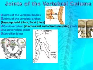
Cartilagenous joints • Amphiarthroses • Restricted movement • Symphyseal joints • Pubic symphysis : bony elements covered by hyaline cartilage with fibrous cartilage in between • Intervertebral discs • Specialised symphysis – hyaline cartilage plates of vertebral bodies joined by ring of fibrous cartilage e.g. annulus fibrosus
Synovial Joints • Diarthroses • Mobile • Articular surfaces covered by hyaline cartilage • Fibrous capsule lined by synovial membrane • Fibrous layer (layers of collagen) of capsule connected to periosteum of bones • Synovial cavity with synovial fluid • Ligaments formed by irregular connective tissue • Menisci / articular discs of fibrous cartilage
Synovial Joints contd • Synovial membrane: • Inner surface smooth / with folds (villi) • Contains blood vessels, lymphatic vessels, nerves • Cells about 1-6 layers on the inside • Type A cells: macrophages • Type B cells: fibroblasts which form synovial fluid
HUMAN DEVELOPMENTTEXTBOOKS • Medical Embryology by J Langman 4th edn • Chapter 9 : Skeletal system p123 • Chapter 10 : Muscular system p138 • The Developing Human by KL Moore 4th edn • Chapter 15 : The skeletal system p334 • Chapter 16 : The muscular system p350
SKELETAL SYSTEM • 3rd wk • Mesoderm – series of blocks = somites • Differentiation • Sclerotome = ventromedial • Dermatome = dorsolateral • 4th wk-sclerotome cells mesenchyme=conn tissue • Migration
SKELETAL SYSTEM • Sclerotome cells migrate • Fibroblasts (matrix formation) • Chondroblasts (cartilage formation) • Osteoblasts (bone formation) • Bone formationMesenchyme • Bone can also form in somatic mesoderm of body wall
LIMBS • 5th wk – limb buds • Initially – mesenchyme core surr by ecto • Apex of bud thickened=ectodermal ridge • Ridge influences underlying mesenchyme • Rapid growth & differentiation • 6th wk – terminal end of buds flat • = hand and foot plates
LIMBS • Tissue disappearance in radial grooves • = fingers & toes • External shape is established • 6th wk – hyaline cartilage model • End of embryonic period • endochondral ossification • 12th wk – primary ossification centers
LIMBS • Ossification progresses from primary center • To the ends • At birth • Diaphysis completely ossified • Epiphyses cartilaginous • Cartilage plate inbetween = epiphyseal plate • Latter NB for growth in length • Full length – plate disappears, epiphyses unite
Clinical application • Fracture vs Normal development • Determination of proper maturation age • Hand and wrists commonly used to determine “bone age”
LIMB ABNORMALITIES • Amelia – 1 or 2 extremities are absent • Meromelia – hands & feet attached to trunk • Micromelia – all extremities present but abnormally short • Polydactyly – extra fingers or toes • Syndactyly – abnormal fusion • Lobster claw – abn cleft betw 2nd & 4th metacarpal (MC) bones & sift tissues; 3rd MC & phalanges almost always absent; thumb, index finger, 4th, 5th finger may be fused. • Club foot – sole in, foot add & plantar flexed