Download
1 / 30
300 likes | 438 Views
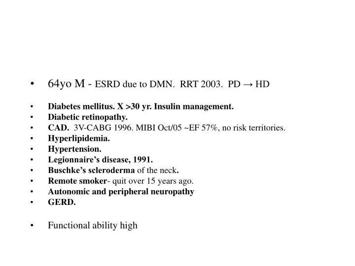
64yo M - ESRD due to DMN. RRT 2003. PD → HD Diabetes mellitus. X >30 yr. Insulin management. Diabetic retinopathy. CAD. 3V-CABG 1996. MIBI Oct/05 ~EF 57%, no risk territories. Hyperlipidemia. Hypertension. Legionnaire’s disease, 1991. Buschke’s scleroderma of the neck .

E N D
64yo M - ESRD due to DMN. RRT 2003. PD → HD • Diabetes mellitus. X >30 yr. Insulin management. • Diabetic retinopathy. • CAD. 3V-CABG 1996. MIBI Oct/05 ~EF 57%, no risk territories. • Hyperlipidemia. • Hypertension. • Legionnaire’s disease, 1991. • Buschke’s scleroderma of the neck. • Remote smoker- quit over 15 years ago. • Autonomic and peripheral neuropathy • GERD. • Functional ability high
Transplant history • Extended Criteria Donor August 10th • Low immunologic risk: Neg PRA & cross-match • Cold Ischemic time 9hr • BENEFIT trial: CsA, MMF & pred. +CsA arm. • DGF: HD on POD #2. • Cr 220umol/L @ d/c POD #7.
U06-15305 #0150171274317 • Donor kidney biopsy
DiagnosisRenal Biopsy: • Hypertensive nephropathy with mild parenchymal atrophy and scarring and less than 20% glomerulosclerosis • Marked tubular degenerative changes. Comment: to have a glomerulus with segmental sclerosis is somewhat more than one would expect to see in the spectrum of hypertensive nephropathy. Whether this could be a sign of primary glomerular disease (early secondary FSGS) should be evaluated clinically.
Transplant history • Early BK rise resulted in progressive reduction in immunosuppression • By Jan/07: MMF 500 bid, Target CsA ~100
marsupilization Bx 3 Bx 1 Bx 2 • 1st Xplant Biopsy Oct 6th. Cr @ 304umol/L. No rejection or BKN. • ?Xplant obstruction –Lymphocele marsupilized Oct 18th. Transient Cr drop to 275umol/L • 2nd Xplant Biopsy Nov 9th for Cr rise 256 → 315umol/L. Rx’d as acute rejection with 3d IV solumedrol –no pred taper. Creatinine 262 umol/L by end November. No BKN • 3rd Xplant Biospy Jan 18th 2007. Creatinine 335 Jan 11, 2007
U06-19146 #622858320 • Renal Tx 10 Aug 06 • Increase serum creatinine • R/O rejection
IF • C4d- Section is folded and hard to interpret. Peritubular capillaries appear negative. The one glomerulus shows some irregular staining which appears to be in the mesangium and around one capillary, interpreted as negative.
DiagnosisRenal Biopsy (2 months post-Tx): • Negative for rejection • Banff scores: • G0 I1 T0 V0 AH1 • Mild to moderate arteriosclerotic small vessel disease, including arteriolar hyalinosis (presumably of donor origin)
marsupilization Bx 3 Bx 1 Bx 2 • 1st Xplant Biopsy Oct 6th. Cr @ 304umol/L. No rejection or BKN. • ?Xplant obstruction –Lymphocele marsupilized Oct 18th. Transient Cr drop to 275umol/L • 2nd Xplant Biopsy Nov 9th for Cr rise 256 → 315umol/L. Rx’d as acute rejection with 3d IV solumedrol –no pred taper. Creatinine 262 umol/L by end November. No BKN • 3rd Xplant Biospy Jan 18th 2007. Creatinine 335 Jan 11, 2007
U06-21520 #622858320 • CAD Tx 3 months ago • Recent surgery to correct urinary obstruction • Recent increase in creatinine • Rejection?
IF • IgG-Negative. • IgA-Negative. • IgM-Negative. • C3- Moderate staining in a small vessel. • C1q-Negative. • Kappa-Negative. • Lambda-Negative. • Fibrinogen- Moderate to strong interstitial staining. • Albumin- Mild non specific background. • C4d- Peritubular capillaries negative. Minimal glomerular staining with a focal and segmental character, mostly in a wispy mesangial distribution.
BK virus ISH • negative
DiagnosisRenal Biopsy (3 months post-transplantation): • Mild tubulo-interstitial rejection • Severe arteriosclerotic vascular disease apparently of donor origin • Banff score: • G0, I2, T2, V0, AH1
marsupilization Bx 3 Bx 1 Bx 2 • 1st Xplant Biopsy Oct 6th. Cr @ 304umol/L. No rejection or BKN. • ?Xplant obstruction –Lymphocele marsupilized Oct 18th. Transient Cr drop to 275umol/L • 2nd Xplant Biopsy Nov 9th for Cr rise 256 → 315umol/L. Rx’d as acute rejection with 3d IV solumedrol –no pred taper. Creatinine 262 umol/L by end November. No BKN • 3rd Xplant Biospy Jan 18th 2007. Creatinine 335 Jan 11, 2007
U07-1117 #622858320 • Transplant in Aug/06 • Lowest creat 220 • Now 330 • BK plasma ++ • Prior biopsy – for BK • +/- rejection on last Nov – 3d steroid – no benefit • D6F • Immunosupp CsA- low levels, cellcept, prednisone
IF • IgG-Negative. • IgA-Negative. • IgM- interstitial plasma cells and lymphocytes. • C3- minimal non specific TBM • C1q-Negative. • Kappa- Occasional interstitial plasma cells. Glomerulus negative. • Lambda- Numerous interstitial lymphocytes or plasma cells. • Fibrinogen- Moderate interstitial staining. • Albumin- Moderate background with some prominence of basement membranes. • C4d- Negative.
IH • EBER negative • Slight predominance of lambda over kappa
DiagnosisRenal Biopsy (5 months post-Tx): • Plasma cell rich acute cellular rejection (tubulointerstitial pattern with a minimal vascular component) • Banff scores • G0 CG0 I3 CI2 T3 CT2 V1 CV2 AH0 MM2
Comment • The obvious feature is tubulointerstitial rejection. • Accompanying vasculitic lesion is minimal and barely sufficient for diagnosis. • Unusual IF: lymphocytes/plasma cells expressing predominantly lambda light chain + IgM • Special studies are not in favor of PTLD • EBV serology (IgM) should be verified