Download
1 / 15
150 likes | 316 Views
“METHODS IN MOLECULAR BIOPHYSICS Structure, Function, Dynamics”. Igor Serdyuk, Nathan Zaccai and J. Zaccai. Cambridge University Press, 2007, 1 12 0 pages. Maxims. C O N T E N T. Classical and advanced methods are included as an equal. Introduction Biophysical methods in the beginning

E N D
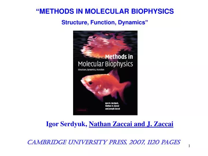
“METHODS IN MOLECULAR BIOPHYSICS Structure, Function, Dynamics” Igor Serdyuk, Nathan Zaccai and J. Zaccai Cambridge University Press, 2007, 1120 pages
Maxims C O N T E N T Classical and advanced methods are included as an equal. Introduction Biophysical methods in the beginning of the 21st century: from ensemble measurements to single molecule detection Part A Biological Macromolecules and Physical Tools Chapter A1 Macromolecules in their environment Chapter A2 Macromolecules as physical particles Chapter A3 Understanding macromolecular structures Part B Mass Spectrometry Chapter B1 Mass and charge Chapter B2 Structure and function studies Part C Thermodynamics Chapter C1 Thermodynamic stability and interactions Chapter C2 Differential scanning calorimetry Chapter C3 Isothermal titration calorimetry Chapter C4 Surface plasmon resonance Part D Hydrodynamics Chapter D1 Macromolecules as hydrodynamic particles Chapter D2 Fundamental theory Chapter D3 Molecular diffusion Chapter D4 Analytical ultracentrifugation Chapter D5 Electrophoresis Chapter D6 Electric birefringence Chapter D7 Flow birefringence Chapter D8 Fluorescence depolarisation Chapter D9 Viscosity Chapter D10 Dynamic light scattering Chapter D11 Fluorescence correlation spectroscopy Part E Optical Spectroscopy Chapter E1 Visible and Infrared absorption spectroscopy Chapter E2 Two-dimensional infrared spectroscopy Chapter E3 Raman scattering spectroscopy Chapter E4 Optical activity Part F Optical Microscopy Chapter F1 Light microscopy Chapter F2 Atomic force microscopy Chapter F3 Fluorescence microscopy Chapter F4 Single molecule detection Chapter F5 Single molecule manipulation Part G X-ray and Neutron Diffraction Chapter G1 Macromolecule as a radiation scattering particle Chapter G2 Small angle scattering Chapter G3 X-ray and Neutron macromolecular crystallography Part H Electron diffraction Chapter H1 Electron microscopy Chapter H2 Images from molecular to atomic resolution Part J Molecular dynamics Chapter J1 Energy and time calculations Chapter J2 Neutron spectroscopy Part K Nuclear magnetic resonance Chapter K1 Frequencies and distances Chapter K2 Experimental techniques Chapter K3 Structure and dynamics studies
Each chapter starts from historical review (each method is deeply rooted) Maxims F2.1 Atomic force microscopy F2.1 Historical review Early 1980s G. Binning and H. Rohrer proposed the scanning tunnelling microscope(STM) (forwhich they were awarded the Nobel Prize). This invention has initiated an excitingseries of novel experiments to image the surface of conducting as well asinsulating solids with atomic resolution. The first attempts at imaging biologicalmolecules using a scanning tunnelling microscope (STM) date back to 1983. In1987, individual molecules of phthalocyanine, lipid bilayers and ascorbic acid were reported. One year later, H. Ohtani with collaborators imaged benzene,the molecule of Kekul´e’s blue dream, as three-lobed rings. In 1989 a spectacularviewof the double-stranded Z-DNAmolecule, the first biological macromolecule studied using a STM, was presented. 1986 G. Binning, C. F. Quate and C. Gerber invented the scanning force microscope(SFM). In this microscope a sensor tip carried by a flexible cantilever is usedto touch and characterise a surface. This was a significant breakthrough which allows biological macromolecules to be scanned in aqueous solution and givesreproducible imaging of DNA and of membrane protein crystals. Early 1990s D. J.Keller,Q. Zhong and C. A. J. Putman independently proposed the so-calledtapping mode of the atomic force microscope (AFM), in which the cantilever isoscillated vertically while it is scanned over the sample. In this mode the imageis formed by displaying the reduction of the oscillation amplitude at every pointof the sample. It was demonstrated that reliable height data for various biologicalmacromolecule assemblies in solution can be obtained using the tapping modeAFM. 2000 to now SFM has become a powerful method for studying thestructure and dynamics of biological macromolecules in solution. Excitingapplications of this technique in biology range from visualisation of transcription in real time to the mechanical manipulation of a single molecule. Essential contributionsto the theory and technique of application of atomic force microscopyto the study of macromolecular structure biological macromolecules were made by C. Bustamante, D. J. Keller, P. K. Hansma, H. G. Hansma, A. Engel andD. J. Müller.
F2. Atomic force microscopy Maxims Each chapter is finished by checklist of key ideas • Atomic force microscopy, also scanning force microscopy, does not use lenses to forman image, but instead uses a sharply pointed sensor, or tip, at the end of a flexiblecantilever to scan and sense the topography of a sample. • The heart of the AFM is a tip mounted on a flexible cantilever; the ideal AFM tip shouldhave a radius that is as small as possible, a well-defined and reproducible molecularstructure and be mechanically and chemically robust such that its structure is not alteredwhile imaging in fluid environments. • In the simplest mode of operation, the contact mode, the AFM resembles a phonograph.As the sample is scanned underneath the tip, the cantilever deflects up or down, trackingthe surface. • In the tapping mode of operation, the cantilever vibrates at its resonance frequency(typically a few hundred kiloherz); the cantilever could be compared to a blind personscanning the environment with a stick to explore the path ahead. • Many factors contribute to an AFM image of biological structures in addition to thetopography of the sample surface;these include the size and shape of the tip,the properties of the feedback loop and the mechanical and chemical properties ofthe sample and imaging environment. • Extraction of submolecular information from AFM topography for isolated globularproteins is a very difficult task; so far it has not been possible to achieve atomic resolutionon soft biological samples. • High-resolution imaging with high signal-to noise ratio in the molecular/submolecularrange can be achieved with two-dimensional crystals of biological macromolecules; AFM enables the process of two-dimensional crystal growth to be visualised in realtime. • It is possible to use the AFM tip like a mechanical ‘nanoscalpel’; after scanning the areaof interest in non-contact mode AFM; biological material (e.g. chromosome bands) canbe cut by the AFM tip at a high force.
Commentary for biologists Maxims Comment D1.2The Reynolds number is a dimensionless parameter, which determines the relative importance of inertial and viscous effects. Bacteria Taking ρ = 1 g cm−3 and η0 =10−2 g cm−1 s−1, we obtained a Reynolds number of 10−5, i.e. very small. The bacterium therefore lives in a world without inertia. Fish The same calculation for a fish of length l = 10 cm, moving with velocity ~ 100 cm s−1 in water yields a Reynolds number of about 105. This is an exampleof hydrodynamics at high Reynolds number. The fish lives in a water medium withinertia. Comment D2.7 Capacitance An electric charge q on a body generates an electrostatic potential, U, proportional to q U = q/C The proportionality constant is 1/C, where C is defined as the capacitance of the body.C has the dimensions of length. For the mathematically minded: C is equal to thecharge required to maintain a body at unit electrostatic potential with respect to infinity or C is equivalent to the electrostatic capacitance of the particle in units inwhich the capacity of a sphere equals its radius. Comment G1.8 Spin incoherence in neutron scattering During a neutron scattering event, a nucleus of spin I combines with a neutron ofspin 1/2 to form one of the two intermediate states I + 1/2 or I -- 1/2, with relativeweights, w+, w−, respectively. Different scattering lengths, b+ and b−, respectively,are associated with each of these states, leading to a total scattering cross-section: σ = S + s = 4π(w+b+ + w−b−) where S is the coherent and s the incoherent part: S = 4π(w+b+ + w−b−)2 s = σ − S Commentary for physicists Comment B2.5Edman degradation In 1950 P. Edman proposed a chemical method forthe stepwise removal of aminoacid residues from the N-terminus of a polypeptide or protein. The series ofreactions has come to be known as the Edman degradation, and the method remainsthe most effective chemical means for polypeptide sequencing. The Edman methodrequires a free amino group at the N-terminus. The peptides in which the terminalamino group is blocked (e.g. by a formyl, acetyl or acyl group) have to be cleaved byeither chemical or enzymatic degradation. Edman degradation is carried out in anautomated analyser. Sequence data can now be obtained from as little as 10-100 ngof protein. Comment F4.6 Peptide nucleic acid Peptide nucleic acid is a polymer in which the phosphate sugar backbone of nucleicacids is replaced by a peptide-like backbone, based on the monomer 2-aminoethylenglycin carrying any of the four nucleobases: A, T, G, C. This polymer, in contrast to RNA or DNA, is electrically neutral. To positively charge it,one could add, in addition to fluorescent label, some lysine or any positively chargedresidues at one of the termini. Comment F4.8 Flavoenzymes “Flavoevzymes are ubiquitous and undergo redox reactions in a reversible manner.Choloesterol oxidase (COx) from Brevibacterium sp. is a 53-kDa flavoenzyme thatcatalyses the oxidation of a cholesterol by oxygen. The active site of the enzymeinvolves FAD, which is naturally fluorescent in its oxidised form but not in itsreduced form. The FAD is first reduced by a cholesterol molecule to FADH2, and isthen oxidised by O2, yielding H2O2. The crystal structure of COx shows that theFAD is non-covalently and tightly bound to the centre of the protein and issurrounded by a hydrophobic binding pocket for cholesterol, which is otherwisefilled with 14 water molecules.
Maxims Conservation of “old gold” results of structural molecular biology FRET as a spectroscopic ruler From α-helix to coil 6M GuHCl is an universal solvent rRNAs are continuous chains Dansyl (Pro)n--Naphthyl poly- γ -benzyl-L-glutamates. Dimethyl formamide Proteins in GuHCl 16S rRNA Dichloracetic acid Stryer and Hougland, 1967 Ch. Tenford, 1968 Doty et al., 1956 Spirin, 1963 Ethanol Haemoglobin Ribosomes DNA Chemical shifting 70S Spin-spin coupling 30S and 50S Tissieres et al., 1958Bloom and Shoolery, 1955R. Franklin and R. Gosling, 1953М. Perutzand J.Kendrew, 1957
State-of-art all biophysical methods Maxims Introduction Biophysical methods in the beginning of the 21st century: from ensemble measurements to single molecule detection Part A Biological Macromolecules and Physical Tools Chapter A1 Macromolecules in their Environment Chapter A2 Macromolecules as physical particles Chapter A3 Understanding macromolecular structures Part B Mass Spectrometry Chapter B1 Mass and charge Chapter B2 Structure and function studies Part C Thermodynamics Chapter C1 Thermodynamic stability and interactions Chapter C2 Differential scanning calorimetry Chapter C3 Isothermal titration calorimetry Chapter C4 Surface plasmon resonance Part D Hydrodynamics Chapter D1 Macromolecules as hydrodynamic particles Chapter D2 Fundamental theory Chapter D3 Molecular diffusion Chapter D4Analytical ultracentrifugation Chapter D5 Electrophoresis Chapter D6 Electric birefringence Chapter D7Flow birefringence Chapter D8 Fluorescence depolarisation Chapter D9 Viscosity Chapter D10 Dynamic light scattering Chapter D11 Fluorescence correlation spectroscopy Part E Optical Spectroscopy Chapter E1 Visible and Infrared absorption spectroscopy Chapter E2 Two-dimensional lnfrared spectroscopy Chapter E3 Raman scattering spectroscopy Chapter E4 Optical activity Part F Optical Microscopy Chapter F1 Light microscopy Chapter F2 Atomic force microscopy Chapter F3 Fluorescence microscopy Chapter F4 Single molecule detection Chapter F5 Single molecule manipulation Part G X-ray and Neutron Diffraction Chapter G1 Macromolecule as a radiation scattering particle Chapter G2 Small angle scattering Chapter G3 X-ray and Neutron macromolecular crystallography Part H Electron diffraction Chapter H1 Electron microscopy Chapter H2 Images from molecular to atomic resolution Part J Molecular dynamics Chapter J1 Energy and time calculations Chapter J2 Neutron spectroscopy Part K Nuclear magnetic resonance Chapter K1 Frequencies and distances Chapter K2 Experimental techniques Chapter K3 Structure and dynamics studies
Analytical centrifugation Mass-spectrometry 70S 5 мМ Мg 5 мМ Мg 1 mM Mg 30S 50S 1 mM Mg Ribosomes in solution Ribosomes in gas phase
Resolution Microscopy 1. Standard light microscopy 2. Near-field scanning microscopy 3. Confocal microscopy 4. Fluorescence microscopy 5. Atomic force microscopy In diffraction limit (250 nm) Out of diffraction limit (70 nm) (60-70 nm) Interior of cells (1-2 nm) Imaging mode 1-10 nm Force-measuring mode
New structural molecular biology requiresnew knowledge Millilitre 10-3 L (m) Microlitre 10-6 L (µ) Nanolitre 10-9 L (n) Picolitre 10-12 L (p) Femtolitre 10-15 L (f) Attolitre 10-18 L (a) Zeptolitre 10-21 L (z) There was no need to know There is the barest necessity
New structural molecular biology requiresnew knowledge Millilitre 10-3 L (m) Microlitre 10-6 L (µ) Nanolitre 10-9 L (n) Picolitre 10-12 L (p) Femtolitre 10-15 L (f) Attolitre 10-18 L (a) Zeptolitre 10-21 L (z) There was no need to know There is the barest necessity
Molecular joga of DNA Natural DNA in different conformations
Molecular joga of DNA Stretching Torsional stress 65 pN B-form P-form B-form S-form
Методы в молекулярной биофизике.Структура, функция, динамика И. Сердюк, Н. Заккаи и Дж. Заккаи Первый том Предисловие Введение: Биофизические методы в начале XXIвека: от измерений вансамбле к детектированию одиночных молекул Часть А. Биологические макромолекулы и физические инструменты Глава A1. Макромолекулы и их окружение Глава A2. Макромолекулы как физические частицы Глава A3. Понимание макромолекулярных структур Часть Б. Масс-спектрометрия Глава Б1. Масса и заряд Глава Б2. Структурно-функциональные исследования Часть В. Термодинамика Глава В1. Термодинамическая стабильность и взаимодействия Глава В2. Дифференциальная сканирующая микрокалориметрия Глава В3. Изотермическая калориметрия титрования Глава В4. Поверхностный плазмонный резонанс Часть Г. Гидродинамика Глава Г1. Биологические макромолекулы как гидродинамические частицы Глава Г2. Фундаментальная теория Глава Г3. Молекулярная диффузия Глава Г4. Аналитическое ультрацентрифугирование Глава Г5. Электрофорез Глава Г6. Ориентация макромолекул электрическим полем Глава Г7. Ориентация макромолекул гидродинамическим полем Глава Г8. Деполяризованная флуоресценция Глава Г9. Вязкость Глава Г10. Динамическое рассеяние света Глава Г11. Флуоресцентная корреляционная спектроскопия
Второй том Часть Д Оптическая микроскопия Глава Д1 Световая микроскопия Глава Д2 Микроскопия силового поля Глава Д3 Флуоресцентная спектроскопия Глава Д4 Детектирование одиночных молекул Глава Д5 Макромолекулярная механика Глава Д6 Биологические моторы Часть Е Рентгеновская и нейтронная дифракция Глава Е1 Макромолекулы как рассеивающие частицы Глава Е2 Малоугловое рассеяние Глава Е1 Рентгеновская и нейтронная кристаллография Часть Ж Электронная дифракция Глава Ж1 Электронная микроскопия Глава Ж2 Трехмерная реконструкция из двухмерных изображений Часть З Оптическая спектроскопия Глава З.1 Спектроскопия в видимой области Глава З.2 Инфракрасная спектроскопия Глава З.3 Двумерная инфракрасная спектроскопия Глава З.4 Рассеяние Мандельштама-Рамана Глава З.5 Оптическая активность с использованием световых и синхротронных источников Часть И Молекулярная динамика Глава И.1 Энергетические и временные вычисления Глава И.2 Нейтронная спектроскопия Часть К Ядерный магнитный резонанс Часть И.1 Частоты и расстояния Часть И.2 Экспериментальная техника Часть И.3 Исследование структуры и динамики


![Cambridge International Dictionary of English [Cambridge University Press] group/gru:p/](https://cdn1.slideserve.com/3307704/slide1-dt.jpg)