Download
1 / 47
500 likes | 919 Views
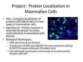
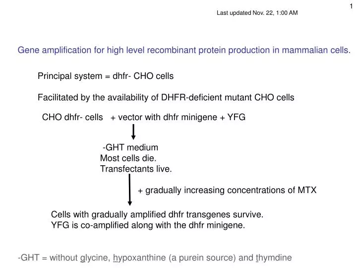
Last updated Nov. 22, 1:00 AM. Gene amplification for high level recombinant protein production in mammalian cells. . Principal system = dhfr- CHO cells Facilitated by the availability of DHFR-deficient mutant CHO cells. CHO dhfr- cells + vector with dhfr minigene + YFG. -GHT medium

E N D
Last updated Nov. 22, 1:00 AM Gene amplification for high level recombinant protein production in mammalian cells. Principal system = dhfr- CHO cells Facilitated by the availability of DHFR-deficient mutant CHO cells CHO dhfr- cells + vector with dhfr minigene + YFG -GHT medium Most cells die. Transfectants live. + gradually increasing concentrations of MTX Cells with gradually amplified dhfr transgenes survive. YFG is co-amplified along with the dhfr minigene. -GHT = without glycine, hypoxanthine (a purein source) and thymdine
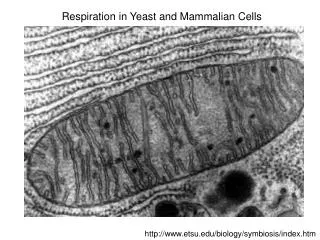
A different major system for high level Mab production • NS0 cells: • Mouse myeloma cells, high IgG producers IgG- variants = NS0 • No endogenous IgG, but cell is a natural IgG secretor. • Lack glutamine synthetase (GS): • glutamate + NH3 + ATP glutamine + ADP + Pi • Vector = MAb genes driven by strong promoters • (2 polypeptides: H-chain and L-chain) • + GS cDNA gene (Bebbington) • Select on glutamine-free medium • Inhibit GS with methionine sulfoximine (gln analog) • Select for GS overproducers • --->--> Gene amplification does not seem to be operating in NS0 cells but can be performed in GS+ CHO cells by suppressing the activity of the endogenous enzyme with methionine sufoximine) • Proprietary (Lonza Biologics)
Resting T- or B-cell) Mature T-cell (effector T-cell) Plasma cell (effector B-cell) Extensive ER Myeloma cells (e.g., NS0) are similar 2002 Molecular Biology of the Cell by Bruce Alberts, Alexander Johnson, Julian Lewis, Martin Raff, Keith Roberts, and Peter Walter.
Transfection strategies for gene amplification • YFG (Your Favorite Gene) linked to a dhfr minigene on a single plasmid A. ~Insures co-integration B. ~Insures co-amplification • YFG and dhfr on separate plasmids A. Allows a high ratio of YFG to dhfr to start B. Co-amplification not assured but commonly occurs.
Amplification protocol Note: Process is lengthy and tedious.
Some marketed recombinant proteins Erythropoietin (Epogen, Procrit) J&J, Amgen Tissue plasminogen activator (TPA) Genentech Growth Hormone (Genentech) Insulin (Genentech) Beta-interferon (Avonex) Biogen-IDEC Alpha-interferon (IntronA) Schering-Plough Neupogen (Amgen) Etanercept – TNF receptor + IgG (Enbrel) Amgen Monoclonal antibodies (mAbs): see next
Ways to increase production and/or lessen development time: Mitchell Reff (IDEC patent): Screen for a high production genomic position. Integrate YFG into it by homologous recombination, selecting for reconstitution of a split dhfr minigene, then amplify. Mitsubishi (T. Shibou, Mitsubishi Pharma Corporation. European Patent Application. Vol. EP001293564A1, PCT/JP01/04801): Same, but use a lox site and site-specific recombination to integrate YFG. Add chromatin remodeling sequences to vector to open chromatin. Add “insulator” sequences to vector to block postion specific repression. Search for even better promoters (current: CMV, EIFalpha, actin) Or even synthetic promoters (E. coli: Stephanopolos, MIT) Engineer cells with advantageous glycosylation patterns Engineer cell to eliminate or defer apoptosis (for longer productinon runs) Etc. (including Chasin lab project)
(2004 claim = 2.8 g/L) Lonza 2005 Web site presentation (1990) ml. Cell titer Antibody grams/liter Ab g/l Millions of cells per liter • Note improvement include: • Higher cell density • Longer times • Higher output per cell (23 d.) (Old values from
20,000 liter fermentor 20,000 liter mammalian cell fermentor - Lonza Biologics - Portsmouth, NH
High level production in mammalian cells. Do the math (back of the envelope): Reff patent (IDEC, now Biogen-Idec): 55 pg/cell/day Max cell density = 107/ml? So 1010 cells/L Therefore 55 x 10-12 g/cell/day x 1010 cells/L = 55 x 10-2 g/L/day = 0.55 g/L/day = 11 g/L/20 days, calculated Lonza (contract manufacturer) claims (2005) = 5 g/L yield Same ballpark. 30,000 L reactor (largest): 30,000 L. X 5 g/L. = 150 kg in 20 days, or say one month x 12 months = 1800 kg/year = ~ 2 tons = 1,800,000 g/year One MAb dose = ~500 mg = 0.5 g 1,800,000/0.5 = 3.6 million doses per reactor per year. 6 doses per patient per year ? 3,600,000/6 = 600,000 patients per year per reactor (market exist?) At $15,000 (low?) per patient per year $9B in sales /per 30 kL reactor
Monoclonal Antibodies (mAbs) • Antibodies (Abs). Also known as immunoglobulins (Ig). • Comprised of 2 heavy chains and 2 light chains • Monoclonal Abs bind specifically to a single site (epitope) on a particular antigen • Abs are produced by B lymphocytes. • Because of their specificity and ease of generation, they are extensively used as therapeutics (“passive immunotherapy”) and as diagnostic and research tools • - They can be generated in large (unlimited) amounts in culture
Antibodies are made by B-cells B cells develop in the bone marrowhematopoietic stem cells and lymphoid stem cells lymphoid stem cells T-cells and B-cells B-cells: progenitor = pro-B cell (B220+) precursor = pre-B cells: heavy-chain rearranged immature B cell: makes IgM + light-chain rearranged matured B cell: Makes IgM + IgD + an antigen encountered in spleen or lymph nodes; then goes to peripheral circulation Terminally differentiated cell = plasma cell, periphery, Ig secretor (IgG, IgM, + some others) Immunocytes at different stages or of different types are often characterized by characteristic specific cell surface proteins, often acting as antigens Each immunocyte (and its offspring) synthesize only a single type of Ig, and use only one of the two alleles available (allelic exclusion) For a summary of the immune system see Strachan and Read, pp. 119-131
} Fab Fragment ,antigen binding } Fc Fragment , crystallizable Domain structure of an immunoglobulin molecule Heavy chain Light chain disulfide bonds C = constant regions V = variable regins (antigen binding) H = heavy chain L = light chain
Heavy chain = blue Light chain = pink
Complementarity determining Region = “CDR” Hypervariable region Ig molecule showing polarity, disulfides, carbohydrate CHO = carbohydrate
Fc functions Opsonization Complement activation Antibody-dependent cell-mediated cytotoxicity (ADCC) Transcytosis Fc Constant region See below
Disulfide bond IgM s J-chain Secretory IgA dimer
Multigene organization of Ig genes –light chains: V, J (variable) and C (constant) –heavy chain: V, D, J, (variable) C (constant) Mechanism of Ab gene rearrangement Recombination signal sequences (RSS)–flank V, D, J gene segments –V-RSS------RSS-D-RSS---------RSS-J
IgGkappa light chain gene rearrangement 76 Vk, 5 Jk, 1 Ck over 2 Mb Light chain genesis DNA + SOMATIC HYPERMUTATION (J) J SPLICING (J) (D,J) SPLICING (D,J) + SOMATIC HYPERMUTATION Heavy chain genesis DNA 95Vh, 30Dh,5Jh, 11Ch over 1.4 Mb L = leader sequence, signal for secretion
Class switch recombination sites Alt. splicing IgD Adapted from Janet Stavnezer http://www.umassmed.edu/faculty/show.cfm?start=0&faculty=300
Choice of constant region exons (class switching) takes place via DNA recombination (below) and alternative splicing of pre-mRNA Sequential recombination can also take place Immunobiology, Charles Janeway, Paul Travers, Mark Walport, Mark Shlomchik
Different constant regions can be chosen via alternative pre-mRNA splicing Immunobiology, Charles Janeway, Paul Travers, Mark Walport, Mark Shlomchik
Alternative splicing within a group of Constant region exons yields two forms of IgM pA pA Developmental Biology, Eighth Edition, Scott F. Gilbert
Fc functions Opsonization: Direct uptake into macrophages of bacteria coated with antibody molecules Complement activation: Activated complement proteins lyse foreeign cells by making holes in their membranes (e.g. bacteria cell membrane) Transcytosis: Antibody-antigen complexes are taken up (endocytosed) on one side of an epithelial cell and directed to the other side, where they are exocytosed Antibody-dependent cell-mediated cytotoxicity (ADCC): Cells with a surface antigen are coated with antibodies that bind via their Fab region. Then killer T-cells use Fc receptors on their surface to recognize the Fc region of the attached bound antibodies and kill them with cytotoxins. Fc
Antibodies can participate in host defense in several ways Also ADCC
ADCC = antibody-dependent cell-mediated cytotoxicity Fc receptor When activated by Fc binding, NK cells release Perforin makes holes in the membrane Granzymes = proteases that initiate apoptosis Fc NK cells = natural killer cells
NK cell Genentech Fc region ADCC = antibody-dependent cell-mediated cytotoxicity
Antibody generation T-cells: cell mediated immune reactions and B-cells: secreted antibodies 2002 Molecular Biology of the Cell by Alberts, Johnson, Lewis, Raff, Roberts, and Walter.
Prexisting B cells that are already producing antibodies that can bind to a specific antigen are stimulated to divide when presented with that antigen. There are many different clones of such precursor cells, each of which is stimulated. The final response is therefore POLYclonal. 2002 Molecular Biology of the Cell by Bruce Alberts, Alexander Johnson, Julian Lewis, Martin Raff, Keith Roberts, and Peter Walter.
The antibody secreting effector cells terminally differentiate (die) but their sister memory cells live on to generate an amplified response upon a second exposure to antigen 1st exposure Effector cells Memory cells 2nd exposure 2002 Molecular Biology of the Cell by Bruce Alberts, Alexander Johnson, Julian Lewis, Martin Raff, Keith Roberts, and Peter Walter.
2002 Molecular Biology of the Cellby Bruce Alberts, Alexander Johnson, Julian Lewis, Martin Raff, Keith Roberts, and Peter Walter.
Monoclonal antibodies via cell hybridization Selects for rare hybrid cells Spleen cells do not grow in culture. TGr myeloma cells do not grow in HAT e.g., in peritoneal cavity) cavity TG = 6-thioguanine HAT = hypoxanthine, amethopterin, and thymidine
Immunize with antigen X Monoclonal antibody generation Hprt- myeloma cells 6-TG-resisatnt HAT- (HAT) Cell fusion Plate among many wells for supernantant testing Selection in HAT Screen for secreted anti-X antibody Plate positives at low density (~1/well) for cloning Positive clones provide a continuing source of anti-X antibody
MAb therapy targets Inflammation Autoimmune disease Graft rejection Cancer Viral infection
Therapeutic strategies • Plain MAbs • MAbs fused to other protein binders (e.g., soluble receptors) to increase avidity and/or to effect ADCC • MAbs fused to cytotoxic agents • (toxins, radionuclides) • Toxins: • ricin (stops protein synthesis) • calicheamicin (DNA breaks) • Radionuclides: • 90Y = yttrium • 111I = indium
Monoclonal antibody generation • - Cells needed myeloma cells and mouse spleen cells • - antigen administration Kohler and Milstein • - hybridoma formation via cell fusion • selection mutants required (myeloma hprt- usually) • Further development: • - antibody generation cDNA cloning from hybridoma • - engineered MAbs expression vectors • refinement 2nd generation antibodies, in vitro • Solve problems of using mouse antibodies in humans
Problems of mouse MAbs • Fc portion limited in its ability to interact with Fc receptors of human cells. • Lower serum half-life • Development of human anti-mouse antibodies (HAMA) • Retreatment results in allergy or anaphylactic shock • Retreatment is less effective Breedveld, Lancet 2000 355:9205 • Solutions via recombinant DNA genetic engineering : • Chimeric mouse-human antibodies: mouse V regions fused to Hu C-region • Humanized mouse antibodies, Parts of V-region from human interspersed with mouse CDR V-regions • Human antibodies (fully), via transgenic mice carrying human immunoglobulin genes as source of spleen cells (Medarex, Abgenix, Kirin) CDR = complementarity-determining region
MAb Fusion Proteins With other protein-binding proteins: natural receptors in soluble form Analogous to MAbs and make use of the Fc portion of the antibody molecule: Example: Enbrel (etanercept): Anti-rheumatoid arthritis drug Soluble TNF receptor fused to the Fc IgG1 domain (TNF= tumor necrosis factor) Ties up TNF, blocking its inflammatory function Fc domain dimerizes the receptor, which increases its affinity for TNF. Fc domain increases the half-life of the protein in the bloodstream Amgen + Wyeth
Example, still experimental: Anti-HIV drug PRO 542 Uses soluble form of the CD4, the molecule to which HIV attaches on T-cells Aim: block the viral surface protein that binds CD4 Soluble CD4 (HIV receptor) fused to IgG2. Tetrameric (all 4 V-regions replaced) – therefore mutlivalent Reduced Fc function (chose IgG2 for this reason) Better half-life than soluble CD4 itself (However, recently replaced by a MAb (PRO 140) targeting the CCR5 cell surface protein, required for viral entry) Progenics
Ag binding site 15 AA linker Single chain antibodies (scFv)
M13 phage display filamentous phage that infects E. coli POI = protein of interest
Phage display to isolate functional V-regions Can be used to screen billions of V-region variants for binding to a particular antigen of choice. Key requirement of this powerful strategy, and many of a similar kind:A physical link of genotype to phenotype 1) a nucleic acid sequence representing a GENOTYPE (here, DNA) to 2) the PHENOTYPE (e.g., binding to something) of a protein coded by that nucleic acid Commonly used phage = m13, filamentous, infects E. coli Phage coat protein the protein the DNA(inside) “Panning” E.g., for a SC Ab, coat the dish with Ag Protein displayed in the phage coatCan screen 1010phage
Phage display selection of scFv’s (single-chain variable regions) Source of sequence: PCR from genome or RT-PCR from mRNA, add randomization (doped synthesis). scFvs Repeat, to reduce background Wash or Elute, re-infect E. coli PCR rescue scFv DNA Clone (plaque on lawn)