Download
1 / 67
720 likes | 1.27k Views
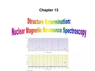
13. Structure Determination: Nuclear Magnetic Resonance Spectroscopy. Why This Chapter? . NMR is the most valuable Used to determine relative location of atoms Maps carbon-hydrogen framework of molecules Depends on very strong magnetic fields

E N D
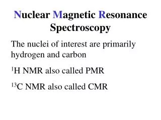
13. Structure Determination: Nuclear Magnetic Resonance Spectroscopy
Why This Chapter? • NMR is the most valuable • Used to determine relative location of atoms • Maps carbon-hydrogen framework of molecules • Depends on very strong magnetic fields • More advanced NMR techniques are used in biological chemistry to study protein structure and folding Mass Spec: Molecular size & formula IR: Functional Groups NMR: Map of Carbons with Hydrogens
13.1 Nuclear Magnetic Resonance Spectroscopy Nuclei w/odd # of protons or odd # of neutrons (~ 1H, 13C, 14N, 2H) spin so act like tiny magnets randomly oriented. Parallel: ~ lower E Antiparallel: ~ higher E external magnet Internal magnetic fields align parallel to or against an aligned external magnet Parallel orientation is lower in energy making this spin state more populated
Nuclear Magnetic Resonance • Radio energy of exactly correct frequency (resonance) causes the parallel nuclei to flip to anti-parallel state Energy needed to flip a spinning parallel nucleus is related to its molecular environment (proportional to field strength, B)
Nuclear Magnetic Resonance • Radio energy of exactly correct frequency (resonance) causes the parallel nuclei to flip to anti-parallel state Energy needed to flip a spinning parallel nucleus is related to its molecular environment (proportional to field strength, B) If nucleus is protected (shielded) from the magnet it takes less E to flip. If nucleus is exposed to the magnet then takes more E to flip.
Example: • I takes 8.0x10⁻⁵ kJ/mol to spin-flip a proton at 200 MHz. Calculate the Energy required to spin-flip a proton (1H) in a spectrometer operating at 300 MHz. 8.0x10⁻⁵ kJ/mol 200 MHz = X kJ/mol 300 MHz X = 1.2x10⁻⁴ kJ/mol
13.2 The Nature of NMR • The sample is dissolved in a solvent that does not have a signal itself Magnet (Applied Field) Radiofrequency energy is transmitted and absorption is detected
The Nature of NMR Absorptions • 1H NMR of methyl acetate has 2 equivalent kinds of H’s so shows 2 peaks • Electrons in neighboring bonds shield or expose nuclei from magnetic field H’s on C next to electron withdrawing C=O H’s on C next to electron withdrawing O • Intensity of 1H NMR peak is proportional to # of equivalent H’s
13C NMR of methyl acetate has 3 kinds of C’s so shows 3 peaks • Electrons in neighboring bonds shield nuclei from magnetic field C next door to electron withdrawing C=O C next to electron withdrawing O C of C=O • Intensity of 13C NMR peak is not related to # of equivalent C’s
At room temperature cyclohexane conformations are interconverting so rapidly that axial and equatorial 1H’s appear identical. • When cold cyclohexane conformations intercovert so slowly that axial and equatorial 1H’s appear different.
Example: • How many signals would you expect each to have in its 1H and 13C spectra? 1H 13C
Solution: • How many signals would you expect each to have in its 1H and 13C spectra? 1H 13C 1 2 3 5
13.3 Chemical Shifts Shift = relative energy of resonance Downfield = deshielded (more exposed to magnet) Upfield = shielded (more protected from magnet) tetramethylsilane [TMS] Reference point
13.3 Chemical Shifts Other signals measured in ppm relative to TMS tetramethylsilane [TMS] Reference point
Measuring Chemical Shift • Numeric value of chemical shift: difference between strength of magnetic field at which the observed nucleus resonates and field strength for resonance of a reference • Difference is very small but can be accurately measured • Taken as a ratio to the total field and multiplied by 106 so the shift is in parts per million (ppm) • Absorptions normally occur downfield of TMS, to the left on the chart • Calibrated on relative scale in delta () scale • Independent of instrument’s field strength
13.4 Signal Averaging & FT-NMR • Carbon-13: (only carbon isotope with a nuclear spin) • Natural abundance =1.1% of C’s so sample is very dilute in this isotope Single run • Sample measured using repeated accumulation of data and averaging of signals, incorporating pulse and the operation of Fourier transform (FT-NMR) • All signals are obtained simultaneously using a broad pulse of energy and resonance recorded • Frequent repeated pulses give many sets of data that are averaged to eliminate noise • Fourier-transform of averaged pulsed data gives spectrum Average of 200 runs
13.5 13C NMR Spectroscopy • Each signal shows different types of environments of carbon • 13C resonances are 0 to 220 ppm downfield from TMS • C’s shift downfield (deshield) if next to electron-withdrawing • Like O, N, X (halogens) sp2 C ~ 110 to 220 sp3 C signal ~ 0 to 90 C(=O) at low field, 160 to 220
Learning Check: Assign resonances in the given 13C spectrum of methyl propanoate 1 2 3 4
Solution: Assign resonances in the given 13C spectrum of methyl propanoate 1 2 3 4
13.6 DEPT 13C NMR • DEPT (distortionless enhancement by polarization transfer) • Normal spectrum shows all C’s then: • Obtain spectrum of all C’s except quaternary (broad band decoupled) • Change pulses to obtain separate information for CH2, CH • Subtraction reveals each type
DEPT 13C NMR Normal spectrum shows all C’s: (Difficult to Assign so many C’s) 5 2 6 DEPT-90: shows only CH’s Quaternary C’s don’t show (Can now narrow our assignments)
DEPT 13C NMR 7,8 Normal spectrum shows all C’s: (Difficult to Assign so many C’s) 5 2 1 6 DEPT-135: Positive =shows CH’s and CH3’s Negative =shows CH2’s (Can narrow assignments even further)
DEPT 13C NMR 7,8 Normal spectrum shows all C’s: (Difficult to Assign so many C’s) 2 5 4 2 1 6 DEPT-135: Positive =shows CH’s and CH3’s Negative =shows CH2’s (Can narrow assignments even further)
13.7 Uses of13C NMR: Example • Evidence for product of elimination of 1-chloro-methyl cyclohexane Expect 5 different C’s; 3 sp3 resonances 20-50 2 sp2resonances 100-150 Expect 7 different C’s; 5 sp3 resonances 20-50 2 sp2resonances 100-150
13.8 1H NMR & Proton Equivalence • Proton NMR is much more sensitive than 13C and the active nucleus (1H) is nearly 100 % of the natural abundance • Shows how many kinds of nonequivalent hydrogens are in a compound • Theoretical equivalence can be predicted by seeing if replacing each H with “X” gives the same or different outcome • Equivalent H’s have the same signal while nonequivalent are different • There are degrees of nonequivalence
Nonequivalent H’s • If replacement of each H with “X” gives a different constitutional isomer then the H’s are in constitutionally heterotopicenvironments and will have different chemical shifts • – they are nonequivalent under all circumstances
Equivalent H’s • Two H’s that are in identical environments (homotopic) have the same NMR signal • Test by replacing each with X if they give the identical result, they are equivalent (homotopic)
Enantiotopic Distinctions • If H’s are in environments that are mirror images of each other, they are enantiotopic • Replacement of each H with X produces a set of enantiomers • The H’s have the same NMR signal (in the absence of chiral materials)
Diastereotopic Distinctions • In a chiral molecule, paired hydrogens can have different environments and different shifts • Replacement of a pro-R hydrogen with X gives a different diastereomer than replacement of the pro-S hydrogen • Diastereotopic hydrogensare distinct chemically and spectrocopically *
Learning Check: • Identify sets of H’s as Unrelated (U), homotopic (H),enantiotopic (E), or diasterotopic (D)
Solution: • Identify sets of H’s as Unrelated (U), homotopic (H),enantiotopic (E), or diasterotopic (D) E D D D D H
13.9 Chemical Shifts in 1H NMR • Proton signals range from 0 to 10 • Electronegative atoms cause downfield shift H’s on sp3C H’s on sp2C Lower field Higher field
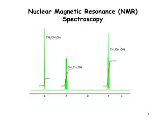
13.10 Integration of 1H NMR Absorptions: Proton Counting • The relative intensity of a signal (integrated area) is proportional to the number of protons causing the signal • For example in ethanol (CH3CH2OH), the signals have the integrated ratio 3:2:1 • For narrow peaks, the heights are the same as the areas and can be measured with a ruler 3 1
13.11 Spin-Spin Splitting in 1H NMR • Peaks are often split into multiple peaks due to interactions between nonequivalent protons on adjacent carbons, called spin-spin splitting • The splitting is into one more peak than the number of H’s on the adjacent carbon (“n+1 rule”) • The relative intensities are in proportion of a binomial distribution and are due to interactions between nuclear spins that can have two possible alignments with respect to the magnetic field • The set of peaks is a multiplet (2 = doublet, 3 = triplet, 4 = quartet)
Simple Spin-Spin Splitting • In bromoethane see 2 kinds of H’s • One at 3.42 and one at 1.68 • Each signal split by neighbors
Simple Spin-Spin Splitting An adjacent CH2 gives a ratio of 1:2:1 An adjacent CH3 group can have four different spin alignments as 1:3:3:1 J (coupling constant) = The separation of peaks in a multiplet is a constant, in Hz
Rules for Spin-Spin Splitting Equivalent protons do not split each other The signal of a proton with n equivalent neighboring H’s is split into n + 1 peaks Protons farther than 2 C’s apart do not split each other
Spin-Spin Splitting Example: • Shift ~1.7 (shielded) • 6 H’s see 1 neighbor (1+1=2 doublet) Integration shows ~6:1 ratio • Shift ~4.3 (deshielded) • 1 H see’s 6 neighbors (6+1=7 septuplet) 6 1
Spin-Spin Splitting Example: Integration shows ratio Triplet ~1.2 (shielded) 3 H’s see 2 neighbors (typical CH3-CH2) Quartet ~2.8 (~deshielded) 2 H’s see 3 neighbors (typical CH3-CH2-c=o) Singlet at 3.8 (deshielded) 3 H’s see 0 neighbors (typical CH3-O) • Shift ~7.8 & 6.8 (deshielded) • 2 H’s see 1 neighbor 2x (1+1=2 doublet) • (typical para pattern) 3 a b 2 3 a b 2 a b 2
Learning Check: From the 1H NMR of C4H10O propose a structure.
Solution: From the 1H NMR of C4H10O propose a structure.
13.12 More Complex Spin-Spin Splitting Patterns: trans-cinnamaldehyde • Spectra more complex if overlapping signals, multiple nonequivalence Doublet of Doublets (dd) ~6.7 (~deshielded) 1 H see’s 2 different neighbors (typical CH-CH-CH) • Doublet ~9.8 (deshielded) • 1 H see 1 neighbor • (typical aldehyde) 1 b 3 a c 3 1 a 1 b 2 c 2 a b 1
13.12 More Complex Spin-Spin Splitting Patterns: trans-cinnamaldehyde • Spectra more complex if overlapping signals, multiple nonequivalence Doublet at 7.5 (deshielded) 1 H see’s 1 neighbor (large J value typical trans alkene H) a = d 2 H’s see 1 neighbor (typical CH-CH) Doublet of Doublets (dd) ~6.7 (~deshielded) 1 H see’s 2 different neighbors (typical CH-CH-CH) • Doublet ~9.8 (deshielded) • 1 H see 1 neighbor • (typical aldehyde) b = dd 2 H’s see 2 different neighbors (typical CH-CH-CH) 1 b 3 a c 3 1 a 1 b 2 c 2 a b 1
13.13 Uses of 1H NMR Spectroscopy • Determine the regiochemistry of hydroboration/oxidation of methylenecyclohexane. Which structure gives this 1H NMR?


![Identification of Stereochemical Isomers of [Mo(CO) 4 (L) 2 ] by Infra-Red Spectroscopy](https://cdn2.slideserve.com/4500730/identification-of-stereochemical-isomers-of-mo-co-4-l-2-by-infra-red-spectroscopy-dt.jpg)