Download
1 / 72
740 likes | 1.04k Views
Neurosensory Disorders: Stroke (CVA, Brain Attack). Marnie Quick RN, MSN, CNRN. A. Pathophysiology/etiology Normal brain physiology and stroke. Ranks 3 rd as cause death Blood supply to one hemisphere is typically blocked, hence terms right & left stroke

E N D
Neurosensory Disorders: Stroke (CVA, Brain Attack) Marnie Quick RN, MSN, CNRN
A. Pathophysiology/etiology Normal brain physiology and stroke • Ranks 3rd as cause death • Blood supply to one hemisphere is typically blocked, hence terms right & left stroke • Functioning brain depends on continuous blood supply for oxygen and glucose & remove end products metabolism
Risk factors for stroke: • Nonmodifible- age, gender, race, family history/heredity • Modifible: hypertension*; atherosclerosis* heart disease; DM; medication (birth control, substance abuse-cocaine/heroin and alcohol; sedetary life style obesity; high cholesterol diet; smoking; stress; sickle cell disease
Brain dysfunction & length of time without blood supply • Brain function depends on collateral circulation and amount of cerebral edema • TIA- neuro deficits last < 24 hrs • RIND- neuro deficits last > 24 hrs but reverse not greater than 21 days • CVA- irreversible brain damage with residual neuro deficits • Stroke-in-evolution- progressive neuro deficits developing over hours or days. Usual cause thrombosis
Ischemic stroke- 80% Occlusion of artery Generally do not lose consciousness Better prognosis than hemorrhagic May have TIA’s before Thrombosis or embolism Hemorrhagic stroke- 15% Bleed occurs with activity Usually rapid onset Generally loss of consciousness Poorer prognosis Intracranial or subarachnoid Two basic disease process causing stroke
Ischemic stroke: Thrombosis • Most common cause of a stroke (60%) • Cause- narrowing of artery from atherosclerotic plaques • Blood is blocked to part of brain that the artery supplies • Often occurs in older individuals who are at rest/sleeping • Tend to form in large arteries that bifurcate, internal carotid artery common site • Can begin as TIA’s, present as stroke-in-evolution, or have completed stroke outright • Lacunar strokes are strokes affecting smaller cerebral vessels in brain- they leave a cavity or ‘lake’
Ischemic stroke Embolism • Caused by: clotted blood from other arteries in the body (heart during atrial fibrillation) fat, bacteria (endocarditis) or air • Emboli circulate until reach an artery in brain that is too narrow to pass through • Usually awake with rapid onset • Extent damage is less severe and recovery faster than other strokes • Will recur if don’t treat cause
Hemorrhagic stroke Intracranial hemorrhage (ICH) • Caused by ruptured artery in the brain • Bleeding varies in size from petechial to massive, edema occurs around the bleed • Blood may form hematoma or be diffuse within the brain • Usually occurs rapidly with the deep arteries • Hypertension is main cause • Most common cause of death due to a stroke • Have more extensive residual deficits and slower recovery than other causes of stroke
Hemorrhagic: Subarchnoid hemorrhage (SAH) • Caused by bleeding into subarchnoid space from • Extension of a intracranial hemorrhage • Aneurysm • AV malformation • Usually occur in younger adults 30-60 than other strokes
Hemorrhagic: SAH Aneurysm • Occur at bifurcations, branches of carotids & vertebrobascular arteries • 85% base brain in anterior circulation • Most common type is berry-bleed from dome • Caused by trauma, congential, arteriosclerosis
Hemorrhagic: SAH- Aneurysm • Aneurysms are graded 0-V on the Hunt/Hess scale; higher the number, poorer chance survival. • Based on LOC & quality of cerebral function • Aneurysm are usually asymptomatic until rupture • Ruptured- sudden explosive headache; loss of consciousness; N & V; nuchal rigidity (stiff neck) and photophobia from meningeal irritation; cranial nerve deficits • Major complications: rebleed, vasospasms, and hydrocephalus
Hemorrhagic: SAH A-V malformation • Congential abnormal joining of arteries to veins in the brain. • As pressures changes occur becomes tangled collection of dilated vessels. • Ischemia symptoms-seizures and interference with normal function of those brain cells
Common manifestations/complications- by body systems
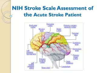
By artery affected by occlusion or hemorrhage Internal carotid
Middle cerebral artery • Contralateral motor loss in the arm and the lower part of the face (central facial palsy) • Contralateral sensory loss in face and arm • Homonymous hemianopsia • Left middle cerebral-communication deficits • Right- spatial/perceptual
Other main arteries off Circle of Willis • Anterior cerebral • Posterior cerebral • Verebrobasilar • Pain or numbness of involved side • Vertigo • Contralateral ataxia • Dysphagia, dysarthria • Cranial nerve dysfunctions
Common Manifestations: Motor deficits • Motor nerve pathways cross in the medulla (brainstem) Prefix hemi- used to describe. Extremities not affected equally- middle cerebral • Amount of motor involvement varies from weakness (-paresis) to paralysis (-plegia). • End paralysis can be flaccid or spastic depending on amount of damage to the motor strip • Initially flaccid and if progress are spastic in 6-8 weeks.
Motor deficits • Facial palsy- (central/UMN) where lower part face affected • Bells palsy (LMN- 7th CN) where the whole side of face affected • Dyphagia- difficulty swallowing
Sensory-perceptual deficits Lack of sensation/propriocetion • Lack of sensation (hemi)- inability to perceive/interpret pain, touch, pressure (post central gyrus) • Lack of/decrease in proprioception or the inability to know where body part is without having to look at it; body’s ‘position sense’
Sensory-perceptual deficits Visual field deficits • Disruption anywhere along the pathway • Homonymous hemianopsia- most common. Loss of half of visual field in each eye. Can’t see toward the same side as the paralysis
Communication Deficits • Motor, speech, language, memory, reasoning, emotions can be affected • Dominant hemisphere for the communication centers is left in most individuals • Global (mixed) aphasia- both expressive (Broca’s area) and receptive (Wernicke’s area) aphasia • Aphasia- total loss of comprehension or use of language • Dysphasia partial loss or difficulty with communication • Dysarthria- difficulty with articulation or muscular control for speech. Sound like have mashed potatoes in their mouth
Communication Deficits Broca’s and Wernicke’s aphasia • Broca’s, expressive or nonfluent aphasia where unable to express- but understands • Wernicke’s, receptive, fluent aphasia- can talk but unable to understand
Communication Deficits Normal process recovery • Begin with one word speech- swearing, ‘ouch’ • Progress to sayings – days of week, social speech, singing • Volitional- normal speech • Recovery may stop at any point, depending on the amount of damage to speech centers
Affect and intellectual deficits • Change level consciousness- confusion to coma • Emotional lability- exaggerated, unpredictable emotional responses. Physiological in nature • Loss of self control, decrease tolerance for stress • Depression, frustration (esp left CVA) • Intellectual changes resulting in memory loss, decreased attention span, poor judgment, inability to think abstractly and make generalizations
Sensory-perceptual deficits Neglect syndrome (unilateral neglect) • Attention disorder in which individual ignores affected part of the body, • Cannot integrate or use perceptions from affected side or from the environment from that side • May observe head turned away from neglected side, does not dress that side, neglects people objects on that side. Diff judging distances • More common in right CVA’s; patient may not be aware of deficit
Inability of the senses to perceive stimuli that were previously familiar. May be any of the senses and varying degrees Inability to carry out purposeful task in the absence of paralysis Or the individual carries out task inappropriately Sensory-perceptual deficits: Agnosia Apraxia
Elimination Deficits • Partial loss of sensation (hemi) can affect perception of need to eliminate bowel/bladder • Cognitive problems may affect the social aspect of elimination • Level of consciousness, immobility, dehydration, diet changes
Immobility complications of Stroke • Any of the immobility complications can occur! • Orthostatic hypotension • Thrombus formation • Impaired respiratory function • Formation of renal calculi • Decreased CO • Osteoporosis • Decubitus ulcer • Contractures
Collaborative Care for Stroke Diagnostic tests • CT- Most important initial- within 25 min Read 45min indicate size location of lesion; differentiate ischemic from hemorrhagic. PET- cerebral blood flow and metabolic activity • MRI or MRA (combined MRI with arteriogram) • Cerebral blood flow • Arteriogram- abnormal structures; vasospasm, stemosis • Transcranial ultrasound doppler velocity of blood flow, degree of occlusion • Cardiac assessment: EKG; cardiac enzymes • Other: LP- obtain CSF, ck bleeding; Blood studies-CBC, glucose, lipid, platelets
Collaborative Preventive care- Manag of modifiable risk factors- medications/surgical • Lewis p1466 Table 58-5 prevention of stroke guidelines and specific treatment for modifiable risk factors with: • medication- treat hypertension, DM, cardiac problems, etc • life style changes: smoking, dietary changes treat hypertension, DM, cardiac problems, etc • Preventative treatment with surgery, such as carotid endarterectomy, stenting
Collaborative Acute Care: Emergency Management of a Stroke • Lewis p. 1468 Table 58-6 • Etiology • Assessment findings • Interventions • Initial • Ongoing
Collaborative Acute Care: Thrombolitic stroke • Medication • Thrombolitic agents to dissolve clot- 3 hrs!!! tPA- Activace. Protocol prior to giving as R/O bleed • Anticoagulants to prevent further extension • Antithrombolitic inhibit platelet phase of clot formation (Ticlid, Plavix, aspirin) • Anticonvulsants prevent seizures from focus • Surgical • Endarterectomy • Angioplasty, carotid artery stenting • Bypass superficial temporal to middle cerebral
Collaborative Acute Care: Embolic/intracranial stroke • Embolic stroke • Medications: If blood clot- anticoagulants, thrombolitic agents, antiarrhythmics; If bacterial- antibiotics • Surgical- Embolic retrieval (Merci retriever) • Intracranial hemorrhage (ICH) stroke • Bedrest • Medication- antihypertensives to normal BP • Surgery- remove hematoma if possible
Collaborative Care: Intracranial Hemorrhage (ICH) • Bedrest • Medication- antihypertensives • Surgical- If hematoma remove
Collaborative Care: SAH • Aneurysm precautions- decrease external/internal stimuli • Medications • Aide with aneurysm precautions- stool softners, antinausea,etc • To prevent rebleed/lysis of clot- Ammicar • To prevent vasospasms- Nimodipine • Before OR- Ca channel blocker- Nimodipine • After OR-triple H- vasodilators (Isuprel); induced arterial hypertension (Dopamine); hypervolemic hemodilution (Albumin) • Prophylactic antiepileptic- Cerebyx/Dilantin
SAH- Common manifestation/complications Major complications • Rebleed due to reabsorption of the clot that is stopping the bleed • Vasospasms due to irritation of the blood vessels • Hydrocephalus from blockage of normal absorption of CSF
SAH A-V malformation • Embolization, ligation of feeders, laser surgery to remove • Gamma Knife- radiation to reduce size of A-V malformation> • Cyberknife below
Collaborative Rehabilitation Care • Physiatrist (rehab physician): Outpatient or in-house rehab • Physical therapy; Occupational therapy; Speech therapy; Cognitive therapy, etc • Exercise program • Outpatient, in-house rehab, nursing home • Home evaluation • Encourage self-care • Community resources • Family support
Nursing assessment specific to stroke Health history & physical exam • Subjective data: Lewis 1472 Table 58-7 • Health history- Risk factors; when symptoms began; describe symptoms; current medications (legal/illegal); other health problems; family history • Objective data • General; respiratory; cardiovascular; GI; urinary; neuro; Vital signs; neuro vital signs (LOC, pupils, motor, sensory) • Level of consciousness- Refer to Module 10