Download
1 / 42
450 likes | 513 Views
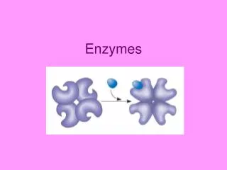
Enzymes: Structure, Properties and Mechanisms of Activity. Factors Affecting Enzyme Activity. In addition to enzyme itself with its cofactors and coenzymes, environmental conditions and enzyme inhibitors may influence on an enzyme activity.

E N D
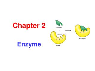
Factors Affecting Enzyme Activity • In addition to enzyme itself with its cofactors and coenzymes, environmental conditions and enzyme inhibitors may influence on an enzyme activity. • Temperature (best be optimum, i.e. the temperature at which enzymatic reaction occur fastest; then high temperatures could be lethal, e.g. denaturation), pH (most like 6 - 8 pH near neutral) and substrate concentration are the main environmental factors. • The rate of reaction increases as substrate concentration increases (at constant enzyme concentration). • Maximum activity occurs when the enzyme is saturated (when all enzymes are binding substrate).
Kinetic properties of enzymes Study of the effect of substrate concentration on the rate of reaction
Effect of enzyme concentration [E] on velocity (v) or reaction rate In fixed, saturating [S], the higher the concentration of enzyme, the greater the initial reaction rate. This relationship will hold as long as there is enough substrate present.
Enzyme Kinetics • Enzyme kinetics deal with determining the rate of a reaction and how it changes in response to changes in experimental parameters. • A key factor affecting the rate of a reaction catalyzed by an E is the concentration of S. Studying the effects of S concentration is complicated by the fact that S changes during the course of an in vitro reaction as S is converted to product. One simplifying approach in kinetic experiments is to measure the initial rate (initial velocity), designated V0. In a typical reaction, the E may be present in nanomolar quantities, whereas S may be five or six orders of magnitude higher. If only the beginning of the reaction is monitored (often the first 60 seconds or less), changes in S therefore can be limited to a few percent, and S can be regarded as constant. • At relatively low concentrations of S, V0 increases almost linearly with an increase in S. At higher S concentrations, V0 increases by smaller and smaller amounts in response to increases in S. Finally, a point is reached beyond which increases in V0 are vanishingly small as S increases. This plateau-like V0 region is close to the maximum velocity, Vmax.
Michaelis and Menten Equation • In 1913, Leonor Michaelis and Maud Menten, developed a kinetic equation to explain the behavior of many simple enzymes. Key to the development of their equation, is the assumption that the E first combines with its S to form an ES complex in a relatively fast reversible step: • k1 • E + S ⇄ ES • k-1 • The ES complex then breaks down in a slower second step to yield the free E and the reaction product P: • k2 • ES ⇄ E + P • k-2 • If the slower second reaction limits the rate of the overall reaction, the overall rate must be proportional to the concentration of the species that reacts in the second step, i.e., ES. • At any given instant in an enzyme-catalyzed reaction, the E exists in two forms, the free or uncombined form E and the combined form ES. At low S, most of the E is in the uncombined E form. Here, the rate is proportional to S because the direction of the first equation above is pushed toward formation of more ES as S increases.
Michaelis and Menten Equation • The maximum initial rate of the catalyzed reaction (Vmax) is observed when virtually all of the E is present in the ES complex and E is vanishingly small. Under these conditions, the E is saturated with its S, so that further increases in S have no effect on rate. This condition exists when S is sufficiently high that essentially all the free E has been converted to the ES form. The saturation effect is a distinguishing characteristic of enzymatic catalysts and is responsible for the plateau observed in figure. The pattern seen in the figure is sometimes referred to as saturation kinetics. • When the E is first mixed with a large excess of S, there is an initial period, the pre-steady state, during which the concentration of ES builds up. This period is usually too short to be easily observed, lasting just microseconds, and is not evident in the figure. The reaction quickly achieves a steady state in which ES remains approximately constant over time. The measured V0 generally reflects the steady state, even though V0 is limited to the early part of the reaction. The analysis of these initial rates is referred to as steady-state kinetics.
Michaelis and Menten Equation • The kinetic curves expressing the relationship between V0 and S have the same general shape (a rectangular hyperbola) for most enzymes, which can be expressed algebraically by the MM equation. Michaelis and Menten derived this equation starting from their basic hypothesis that the rate-limiting step in enzymatic reactions is the breakdown of the ES complex to product and free enzyme. The MM equation is: V0 = Vmax[S]/(Km + [S]). • All these terms, [S], V0, Vmax, as well as the constant called the Michaelis constant, Km, can be readily measured experimentally. • The MM equation describes the kinetic behavior of a great many enzymes, and all enzymes that exhibit a hyperbolic dependence of V0 on S are said to follow Michaelis-Menten kinetics. However the MM equation does not depend on the relatively simple two-step reaction mechanism discussed above. Many enzymes that follow MM kinetics have quite different mechanisms, and enzymes that catalyze reactions with six or eight identifiable steps often exhibit the same steady-state kinetic behavior. Even though the MM equation holds true for many enzymes, both the magnitude and the real meaning of Vmax and Km can differ from one enzyme to another. This is an important limitation of the steady-state approach to enzyme kinetics.
Michaelis and Menten Equation • An important numerical relationship emerges from the MM equation in the special case when V0 is exactly one-half Vmax. Km is equivalent to the substrate concentration at which V0 is one-half Vmax. • The Km can vary greatly from enzyme to enzyme, and even for different substrates of the same enzyme. The Km is sometimes used (often inappropriately) as an indicator of the affinity of an enzyme for its substrate. Thus Km cannot always be considered a simple measure of the affinity of an enzyme for its substrate. • The meaning of the quantity Vmax also varies greatly from one enzyme to the next.
Enzyme Regulation • Biochemical pathways in the living organisms need sophisticated mechanisms for their regulation, due to several reasons: • 1- Maintenance of an ordered state (i.e. timely production of substances without wasting substances). • 2- Conservation of energy (regulating the level of energy-generating reactions just enough to meet the energy requirements). • 3- Responsiveness to environmental changes (regulating the rate of specific reactions to enable cells make relatively rapid adjustments to variations in Temp, pH, ions, etc). • Adjustment of the concentration and activities of certain enzymes is key to the regulation of biochemical pathways. Control of enzymes concentration and activities is accomplished by one of the followings: • 1- Genetic control. Enzyme induction (i.e. the synthesis of enzymes in response to changing metabolic need) is an efficient way of response of cell to changes in environment. Enzyme repression (inhibition of synthesis of certain key enzymes) may be accomplished by the end product of a biochemical pathway.
Enzyme Regulation • 2- Covalent modification. It is the regulation by reversible interconversion between an active and inactive forms of the enzyme molecule due to covalent modifications of enzymes structure.Covalent attachment of a molecule to an amino acid side chain of a protein can modify activity of enzyme. • Many such enzymes have specific residues that may be phosphorylated and dephosphorylated, methylated and demethylated, acetylated and deacetylated or adenylated (the covalent addition of the nucleotide adenosine monophosphate) and deadenylated. 3- Allosteric regulation. In each biochemical pathway at least one enzyme sets the rate for the entire pathway (i.e. pacemaker or regulatory enzyme). This enzyme usually catalyzes the first unique or committed step in the pathway. Another typical control point is the first step of a branch in a pathway that leads to an alternate product. Both covalent modification and allosteric regulation are capable of regulating pacemaker enzymes. Cells use allosteric regulation to respond effectively to changes in intercellular conditions.
Enzyme Regulation Allosteric enzymes are usually composed of several promoters whose properties are affected by effector molecules. Allosteric enzymes have a second regulatory site(allosteric site) distinct from the active site. Allosteric enzymes contain more than one polypeptide chain (have quaternary structure). Allosteric modulatorsbind noncovalently to allosteric site and regulate enzyme activity via conformational changes. • The binding of an effector (ligands) to an allosteric enzyme can affect the binding of substrate to that enzyme. Allosteric effects may be positive or negative. The binding of an effector shifts the curve (enzyme activity in response to S concentration) to a higher (i.e. left or decrease in Km: activator) or lower (i.e. right or increase in Km: inhibitor) activity. Positive modulator binds to the allosteric site and stimulates activity. Positive modulator of an enzyme usually is the substrate of the reaction. Negative feedback inhibition is a process in which the product of a pathway inhibits the activity of the pacemaker enzyme. Negative modulator (inhibitor) binds to the allosteric site and inhibits the action of the enzyme. Usually it is the end product of a biosynthetic pathway (i.e. end-product inhibition).
Enzyme Regulation • Example: Phosphofructokinase (catalyzes the transfer of a phosphate group from ATP to the OH group on C-1 of fructose-6-phosphate) is the main regulatory control point in glycolysis. The enzyme is stimulated by ADP, AMP and other metabolites and inhibited by PEP, citrate and ATP. ATP is a S if binds to active site but is an inhibitor if binds to the allosteric site of the enzyme. • 4- Compartmentation. In eukaryotic cells biochemical pathways are segregated into different organelles. Main purpose of this physical separartion is that opposing processes are easier to control in this way. E.g. FA biosynthesis occurs in the cytoplasm but FA oxidation during energy generation occur in mitochondria. Another purpose of the compartmentation is that each organelle can concentrate specific substances such as substrates and coenzymes. The third purpose is that special microenvironments are often created within organelles. E.g. lysosomes contain hydrolytic enzymes mainly because these enzymes require a high concentration of hydrogen ions for optimum activity (lysosome pH = 5 vs cytoplasm pH = 7.2).
Example of allosteric enzyme - phosphofructokinase-1 (PFK-1) • PFK-1 catalyzes an early step in glycolysis • Phosphoenol pyruvate (PEP), an intermediate near the end of the pathway is an allosteric inhibitor of PFK-1 PEP
Dephosphorylation reaction Usually phosphorylated enzymes are active. Enzymes taking part in phospho-rylation are called protein kinases Enzymes taking part in dephosphorylation are called phosphatases
Reversible and Irreversible Inhibitors Reversibleinhibitors – after combining with enzyme (EI complex is formed) can rapidly dissociate. EI complex is held together by weak, noncovalent interaction. Enzyme is inactive only when bound to inhibitor. Reversible inhibition could be competitive or non-competitive. Competitive inhibitor has a structure similar to the substrate thus can bind to the same active site. The enzyme cannot differentiate between the two compounds. When inhibitor binds, prevents the substrate from binding. Inhibitor can be released by increasing substrate concentration. Non-competitive inhibitor binds to an enzyme site different from the active site. Inhibitor and substrate can bind enzyme at the same time. Cannot be overcome by increasing the substrate concentration. Suicide inhibitor. Inhibitor binds as a substrate and is initially processed by the normal catalytic mechanism. It then generates a chemically reactive intermediate that inactivates the enzyme through covalent modification. It is called suicide because enzyme participates in its own irreversible inhibition.
Multienzyme Complexes and Multifunctional Enzymes • Multienzyme complexes: different enzymes that catalyze sequential reactions in the same pathway are bound together. • Multifunctional enzymes: different activities may be found on a single, multifunctional polypeptide chain. • Metabolite channeling:is “channeling” of reactants between active sites. It occurs when the product of one reaction is transferred directly to the next active site without entering the bulk solvent. It can greatly increase rate of a reaction. • Channeling is possible in multienzyme complexes and multifunctional enzymes. • Metabolism: is the entire network of chemical reactions carried out by living cells. Metabolism also includes coordination, regulation and energy requirement. • Metabolites: are small molecule intermediates in the degradation and synthesis of polymers. • Most organism use the same general pathway for extraction and utilization of energy. All living organisms are divided into two major classes of autotrophs and heterotrophs.
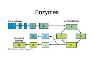
Metabolic Pathways A sequence of reactions that has a specific purpose (for instance: degradation of glucose, synthesis of fatty acids) is called a metabolic pathway. Metabolic pathway may be: (c) Spiral pathway (fatty acid biosynthesis) (a) Linear (b) Cyclic
Catabolism and Anabolism Metabolic pathways can be grouped into two paths: catabolismandanabolism. Catabolic reactions - degrademolecules to create smaller molecules and energy. Anabolic reactions - synthesize molecules for cell maintenance, growth and reproduction. Catabolism is characterized by oxidation reactions and by release of free energy which is transformed to ATP. Anabolism is characterized by reduction reactions and by utilization of energy accumulated in ATP molecules. • Catabolism and anabolismare tightly linked together by their coordinated energy requirements: catabolic processes release the energy from food and collect it in the ATP; anabolic processes use the free energy stored in ATP to perform work. • Metabolism is highlyregulated to permit organisms to respond to changing conditions. Most pathways are irreversible. • Flux - flow of material through a metabolic pathway which depends upon: (1) Supply of substrates (2) Removal of products (3) Pathway enzyme activities
Feedback inhibition • Product of a pathway controls the rate of its own synthesis by inhibiting an early step (usually the first “committed” step (unique to the pathway) Feed-forward activation • Metabolite early in the pathway activates an enzyme further down the pathway
Stages of metabolism Catabolism Stage I. Breakdown of macromolecules (proteins, carbohydrates and lipids to respective building blocks. Stage II. Amino acids, fatty acids and glucose are oxidized to common metabolite (acetyl CoA) Stage III. Acetyl CoA is oxidized in citric acid cycle to CO2 and water. As result reduced cofactor, NADH2 and FADH2, are formed which give up their electrons. Electrons are transported via the tissue respiration chain and released energy is coupled directly to ATP synthesis.
Glycerol Catabolism
Catabolism is characterized by convergence of three major routs toward a final common pathway. Different proteins, fats and carbohydrates enter the same pathway – tricarboxylic acid cycle. Anabolism can also be divided into stages, however the anabolic pathways are characterized by divergence. Monosaccharide synthesis begin with CO2, oxaloacetate, pyruvate or lactate.Amino acids are synthesized from acetyl CoA, pyruvate or keto acids of Krebs cycle. Fatty acids are constructed from acetyl CoA. On the next stage monosaccharides, amino acids and fatty acids are used for the synthesis of polysaccharides, proteins and fats.
Compartmentation of Metabolic Processes in Cell • Compartmentation of metabolic processes permits: • - separate pools of metabolites within a cell • - simultaneous operation of opposing metabolic paths • - high local concentrations of metabolites • Example: fatty acid synthesis enzymes (cytosol), fatty acid breakdown enzymes (mitochondria)
OXIDATIVE DECARBOXYLATION OF PYRUVATE Pyruvate formed in the aerobic conditions undergoes conversion to acetyl CoA by pyruvate dehydrogenase complex. Pyruvate dehydrogenase complex is a bridge between glycolysis and aerobic metabolism – citric acid cycle. Pyruvate dehydrogenase complex and enzymes of cytric acid cycle are located in the matrix of mitochondria.
Entry of Pyruvate into the Mitochondrion Pyruvate freely diffuses through the outer membrane of mitochon-dria through the channels formed by transmembrane proteins porins. Pyruvate translocase, protein embedded into the inner membrane, transports pyruvate from the intermembrane space into the matrix in symport with H+ and exchange (antiport) for OH-.
Conversion of Pyruvate to Acetyl CoA • Pyruvate dehydrogenase complex (PDH complex) is a multienzyme complex containing 3 enzymes, 5 coenzymes and other proteins. Pyruvate dehydrogenase complex is giant, with molecular mass ranging from 4 to 10 million daltons. Electron micrograph of the pyruvate dehydrogenase complex from E. coli.
Enzymes: E1 = pyruvate dehydrogenase E2 = dihydrolipoyl acetyltransferase E3 = dihydrolipoyl dehydrogenase Coenzymes: TPP (thiamine pyrophosphate), lipoamide, HS-CoA, FAD+, NAD+. TPP is a prosthetic group of E1; lipoamide is a prosthetic group of E2; and FAD is a prosthetic group of E3. The building block ofTPP is vitamin B1 (thiamin); NAD – vitamin B5(nicotinamide); FAD – vitamin B2 (riboflavin), HS-CoA – vitamin B3 (pantothenic acid), lipoamide – lipoic acid
Pyruvate dehydrogenase complex is a classic example of multienzyme complex Overall reaction of pyruvate dehydrogenase complex The oxidative decarboxylation of pyruvate catalized by pyruvate dehydrogenase complex occurs in five steps.
Synthesis of glycogen Glucose Pentose phosphate pathway Glucose-6-phosphate Glycogen Ribose, NADPH Degradation of glycogen Gluconeogenesis Glycolysis Ethanol Lactate Pyruvate Fatty Acids Amino Acids Acetyl Co A The citric acid cycle is the final common pathway for the oxidation of fuel molecules — amino acids, fatty acids, and carbohydrates. Most fuel molecules enter the cycle as acetyl coenzyme A.
citrate synthase 1. Citrate Synthase • Citrate formed from acetyl CoA and oxaloacetate • Only cycle reaction with C-C bond formation • Addition of C2 unit (acetyl) to the keto double bond of C4 acid, oxaloacetate, to produce C6 compound, citrate
2. Aconitase aconitase aconitase • Elimination of H2O from citrate to form C=C bond of cis-aconitate • Stereospecific addition of H2O to cis-aconitate to form isocitrate
3. Isocitrate Dehydrogenase isocitrate dehydrogenase isocitrate dehydrogenase • Oxidative decarboxylation of isocitrate toa-ketoglutarate (a metabolically irreversible reaction) • One of four oxidation-reduction reactions of the cycle • Hydride ion from the C-2 of isocitrate is transferred to NAD+ to form NADH • Oxalosuccinate is decarboxylated to a-ketoglutarate
a-ketoglutarate dehydrogenase 4. The -Ketoglutarate Dehydrogenase Complex • Similar to pyruvate dehydrogenase complex • Same coenzymes, identical mechanisms • E1 - a-ketoglutarate dehydrogenase (with TPP) E2 – dihydrolipoyl succinyltransferase (with flexible lipoamide prosthetic group) E3 - dihydrolipoyl dehydrogenase(with FAD)
5. Succinyl-CoA Synthetase Succinyl-CoA Synthetase HS- + GTP + ADP GDP + ATP • Free energy in thioester bond of succinyl CoA is conserved as GTP or ATP in higher animals (or ATP in plants, some bacteria) • Substrate level phosphorylation reaction
Succinate Dehydrogenase 6. The Succinate Dehydrogenase Complex • Complex of several polypeptides, an FAD prosthetic group and iron-sulfur clusters • Embedded in the inner mitochondrial membrane • Electrons are transferred from succinate to FAD and then to ubiquinone (Q) in electron transport chain • Dehydrogenation is stereospecific; only the trans isomer is formed
7. Fumarase Fumarase • Stereospecific trans addition of water to the double bond of fumarate to form L-malate • Only the L isomer of malate is formed
Malate Dehydrogenase 8. Malate Dehydrogenase Malate is oxidized to form oxaloacetate.