Download
1 / 22
220 likes | 333 Views
Journal Club March 2012 A Visual Presentation. Brain Reorganisation and Upper Limb Recovery. Objectives:. Hmm…The elephant in the room, perhaps? Can we be certain that our stroke rehabilitation efforts are achieving better outcomes than if we did nothing at all (natural history)?.

E N D
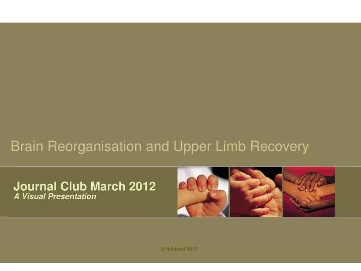
Journal Club March 2012A Visual Presentation Brain Reorganisation and Upper Limb Recovery IJ Hubbard 2012
Objectives: Hmm…The elephant in the room, perhaps? Can we be certain that our stroke rehabilitation efforts are achieving better outcomes than if we did nothing at all (natural history)? • Brain Reorganisation • What’s it all about? • Neuroanatomy update • Brief review of brain topography • Stroke: the disconnect • What’s happened and why? • Upper limb (UL) recovery • Answering the all-important ‘so what’ question This presentation is for your eyes only! Work your way through the 22 slides, take the suggested detours, & then let’s chat! Note from Isobel: IJ Hubbard 2012
Presenter: • Ms Isobel Hubbard • Occupational Therapist working as an Academic Researcher • Lecturer at the University of Newcastle • Convenor of a stroke-specific Masters and Graduate Certificate • Coordinator of all stroke-specific postgraduate & undergraduate courses • PhD Student • Investigating early brain reorganisation & UL recovery post stroke • Track record in publications & presentations Acknowledging the contribution of Drs Parsons, Carey and Budd, the acute stroke team at John Hunter hospital, Newcastle Australia, the National Stroke Foundation and the University of Newcastle’s Priority Research Centre for Brain & Mental Health IJ Hubbard 2012
Brain Reorganisation Definition: Wiki plus IH: Neuroplasticity refers to the ability of the brain and nervous system.. to change structurally and functionally as a result of input from the environment. Plasticity occurs on a variety of levels, ranging from cellular changes involved in learning, to large-scale changes involved in cortical remapping in response to injury, eg stroke...During most of the 20th century, the general consensus.. was that the brain’s structure is relatively stable after a developmental period in early childhood. This belief has been challenged by new findings revealing that the brain is relatively plastic and in turn, responsive to change, throughout an adult’s lifetime. We know from Elbert et al’s research (1995) that if you decided to learn to play the violin, your brain would increase the cortical area allocated to your non-dominant UL Source: wikipedia.org Ref:T Elbert, C Pantev, C Wienbruch, B Rockstroh, E Taub. (1995) Increased cortical representation of the fingers of the left hand in string players. Science 270, 305-307. IJ Hubbard 2012
Brain Reorganisation To find out more: Go to Rossini, Calautti, Pauri & Baron (2002) Post stroke plastic reorganisation in the adult brain. Lancet Neurology, 2, 439-502 Quote: “Our understanding of the mechanisms that promote or prevent recovery is fundamental to the design of novel therapies…In this review we discuss brain imaging studies of reorganisation after stroke. Because motor function of the arms can be used to model the recovery of higher functions…we will focus on the recovery of the sensorimotor arm and hand function. Carey & Seitz (2007) Functional neuroimaging in stroke recovery and neurorehabilitation: conceptual issues and perspectives. International Journal of Stroke, 2 (4), 245-264 Quote: “Our current mandate is to adopt a more active approach to rehabilitation by developing approaches that aim to restore underlying capacity and improve clinical outcome…Promising approaches need to be systematically tested..” • Brain reorganisation: a neural mechanism that underpins stroke recovery because of its responsiveness to changes in behavioural demands • Functional Magnetic Resonance Imaging (fMRI): a non-invasive neuroimaging technique that provides a “window” into brain reorganisation IJ Hubbard 2012
Neuroanatomy 101 Inter-hemispheric fissure: hemispheres are similar but not exact opposites. • The surface of both hemispheres is made up of many gyri (ridges) & sulci (creases). • The Cortex: ≈ top 1cm of gyri and sulci • The Subcortex: the supporting structure • The Cerebellum, Brain Stem & Pons are positioned to connect the brain & body. The central sulci separates the: Pre-central and post-central gyri. Cerebellum The human brain uses up 20% of the body’s oxygen & nutrients • Before you go to the next slide, test your knowledge: • Which regions of the brain are important to motor function and where are they? • Where do you find the cingulate area and what is its primary function? • What % of the corticospinal tracts arising from • the right primary motor cortex (M1) • go to the left UL? IJ Hubbard 2012
Neuroanatomy 101 Left Hemisphere: Brodmanns Area (BA) Functional Regions of Interest BA4: Primary motor cortex (M1) BA6: Premotor cortex (PMC) & Supplementary motor area (SMA) BA3,1,2: Primary somatosensory cortex BA5 & 7: Secondary somatosensory Notes: The SMA, an area important to UL recovery post stroke, is positioned in the medial region of each hemispere (not shown). The two SMA “face-off” across the interhemispheric fissure. Do they communicate via the anterior commissure, a bundle of nerve fibres connecting the hemispheres? To explore the imaged brain: Go to The Whole Brain Atlas at www.med.harvard.edu/aanlib/home.html Select: “NEW: normal anatomy in 3D..”. Use the arrows to navigate the imaged “slices” & try the ‘stroke’ links. Right hemisphere: similar but not exact mirror image Central Sulci Pareital lobe Frontal lobe Source: wikipedia.org Temporal lobe IJ Hubbard 2012
Neuroanatomy 101 • Neural System: a complex labyrinth • Cortex: The end point or journey destination • Subcortex: The connection, journey or road map • Notes re: The brain’s neural system… • Communicates inter- (between) & intra- (within) hemispherically. Lots of connectivity! • Is connected to the body via corticospinal tracts • ≤90% contralateral (opposite side) • ≥10% ispsilateral (same side) • Is dependent on the vascular system for a reliable supply of oxygen & nutrients The brain’s neural system is like a city’s road network Source: wikipedia.org IJ Hubbard 2012
To appreciate the complexity of the neural system go to this youtube link. You’re looking at tractography or connectivity mapping. The different colours represent different directions of activity along the tracts. Interhemispheric connectivity The human brain makes up only 2% of body weight, yet it accounts for 20% of the body’s total O2 consumption & receives 11% of its cardiac output (Giedde, 2006) Neuroanatomy 101 Ref: Gjedde A. Brain energy metabolism and the physiological response, in Functional MRI: An introduction to methods, Jezzard, Mathews & Smith, Ed. 2006, Oxford University Press http://www.youtube.com/watch?v=hUZeuzXH-zA http://en.wikipedia.org/wiki/Diffusion_MRI IJ Hubbard 2012
Neuroanatomy 101 The brain’s vascular system is like its trains & underground • Vascular System: a supply line • Middle Cerebral artery (MCA): Supplies much of the brain’s motor-related neural regions • Circle of Willis: The largest of the brain’s unique contralateral/collateral supply lines • Notes: • This system is responsible for a reliable supply of O2 & nutrients to the brain • A disruption to the blood supply impacts the neural system: Time is brain! • Hyper-acute stroke management is about restoring the integrity of the vascular system ASAP!! FAST!! Time is brain! Each minute 1.9 million neurons die without O2 & nutrients Saver, J.L. (2005) Time is brain – quantified. Stroke 2006 37: 10 IJ Hubbard 2012
To find out more: Go to The Internet Stroke Centre http://www.strokecenter.org/ and you’ll find this image and lots more Neuroanatomy 101 IJ Hubbard 2012
Stroke: the disconnect Hmm…The day after a stroke am I right to assume there’s nothing wrong with the UL muscles? • Stroke is a disconnection phenomena • The integrity of the ‘up-stream’ neural regions • is immediately threatened after any disruption • to the vascular supply, such as stroke. • Ischemic Stroke: Blockage: 80-85% of all strokes • Results in permanent damage ‘up-stream’ • Can present as a squid-like area of damage that’s reached into closely located gyri • Hemorrhagic Stroke: Bleed:15-20% of all strokes • More easily identified via CT scan • Also results in up-stream damage • More life-threatening Time is Brain Act FAST: Face, Arm, Speech & Time http://www.youtube.com/watch?v=T_CXqfFGpvY&feature=relmfu http://www.strokefoundation.com.au/ If you’re unsure about recognising stroke and/or what to do, go to either of these links IJ Hubbard 2012
Predicting Recovery Another elephant…? Should therapists be spending their very limited time on stroke patients who are unlikely to recover? Predicting good vs poor recovery potential • Predicting Recovery: Acute therapists need to be able to quickly & accurately predict potential. • Notes: Strokes that affect the UL.. • Are usually an Anterior Circulation Infarct (ACI) • Have usually impacted the pre- and/or post-central gyri or sensorimotor ‘strip’. • Ref: Oxfordshire Classification system: http://www.strokecentre.org/trials/scales/oxford.html • Stroke topography: Imaging results assist in identifying • where a stroke is located. This is an important indicator of • recovery potential: • Poorest potential: TACI (total) • Best potential: Lacuna infarct (LACI) • What about corticospinal tract integrity? • Ref: Stinear et al (2007) Functional • potential in chronic stroke patients depends on corticospinal tract integrity. Brain, 130, 170-180 Those with ≥20°movement at the wrist & ≥10° in at least one finger To find out more about the corticospinal tract: Go to The Brain from Top to Bottom athttp://thebrain.mcgill.ca/flash/i/i_06/i_06_cl/i_06_cl_mou/i_06_cl_mou.html IJ Hubbard 2012
Before you move onto the next slide take some time-out to consider & write down your 4 most important “best practice” principals for post stroke upper limb rehabilitation. Upper Limb Recovery IJ Hubbard 2012
Upper Limb Recovery Hmm..a room of elephants..!!! Emerging evidence tells us that hospital & rehabilitation wards are usually environmentally bereft and occupationally de-challenging, perhaps making them the worst spaces for people recovering from stroke!! Time is function! In the first month after stroke each day spent doing very little is a lost opportunity for better recovery Four “Most Important” Principals (IH) As Early As Possible: As soon as the patient is medically stable As Intense As Possible: As intense as the patient can reasonably tolerate, which can only be achieved if it’s mostly self-directed As Client-centred As Possible: As meaningful as is environmentally possible As Task-specific As Possible: As close to real-life activities as is feasible and reasonably safe, which is only possible if we take some risks Ref: Hubbard.et al (2009) Task-specific training: Evidence for and translation to clinical practice. Occupational Therapy International, 16(3-4), 175-189 IJ Hubbard 2012
Upper Limb RecoveryUnderstanding the research Typical research question: Do different intensities of UL intervention result in different patterns of activation? • We asked this question & we’re finding differences in certain regions - we’re in the process of writing this up. • Do you find it difficult to understand the reporting of research • Investigating changes in brain activation? Terms often used: • Contralesional: the stroke-affected (lesioned) hemisphere • Ipsilesional: the non-affected hemisphere • Researchers will use analysis methods such as “flipping” the • hemispheres of one group of study participants, so that they can • compare results between hemispheres – as we are doing. • 3. Laterality Index (LI): Shifts in activation • patterns between hemispheres over time – • another way of reporting changes in activation Glass Maps The image above shows the between-group differences in activation over a nominated p-value threshold. SPM is the program used to do the analysis IJ Hubbard 2012
Upper Limb RecoveryWhat’s the research telling us? All systematic reviews found activation associated with the stroke-affected UL was in ipsilesional, motor-related areas eg, the M1 & SMA. IJ Hubbard 2012
Upper Limb RecoveryGood versus Poor Recoverers Important research question: Are there differences in activation patterns between good and poor recoverers? Dr Leeanne Carey et al (2006) Evolution of Brain Activation with good and poor motor recovery after stroke. Neurorehab & Neural Repair, 20, 24-41. Participants: n=9; baseline 2-7 wks post stroke; follow-up at 6 months Method: Compared to healthy volunteer data Findings:Good recoverers with moderate impairment (n=5) had ipsilesional SM1 activation, bilateral SMA & contralesional PMC; the bilateral SMA activation reduced over time. Poor recoverers with severe impairment (n=4) had limited ipsilesional S1M1 and SMA activation & no significant change over time. Conclusion: In the sub-acute phase, there is evidence of reversion to more normal patterns in good recoverers over time. SMA is important to UL recovery following stroke. SMA IJ Hubbard 2012
Joining up the dots….. Knowing what activation patterns are associated with good recovery means researchers can investigate which UL interventions elicit those patterns Upper Limb Recovery IJ Hubbard 2012
Upper Limb RecoveryThe “hopping” problem Time is function! Adequate dose should be a primary consideration irrespective of time post stroke Post Stroke : Upper Limb vs Lower Limb Lower limb: Fortunately it’s more difficult to hop than walk, therefore learned non-use is not an issue. Upper Limb: Unfortunately it’s quite easy to learn to do things one-handed so learned non-use is a big problem that is best challenged ASAP! Constraint-based Intervention: Forces the person to stop UL “hopping”! It challenges learned non-use by forcing the engagement of the stroke-affected UL & demands involvement from both hemispheres. Not suitable for those with no observable movement. Behavioural Demands: Brain reorganisation is a normal response to behavioural changes. Learn the violin and the brain reorganises! Have a stroke and the brain reorganises! Start to walk, speak and/or use the affected UL and the brain reorganises. IJ Hubbard 2012
In Conclusion • Evidence concerning post stroke brain • activation patterns tells us that: • The brain can reorganise post stroke • Brain reorganisation underpins sub-acute • recovery • Our selection of UL interventions should • consider their impact on brain activation • Best practice for most stroke-affected • upper limbs is early more intensive, • task-specific intervention In 2011 we built ourselves a new house with an upstairs and downstairs area. Q: Have we lost sight of the fact that stroke is an “upstairs” disconnection, because our UL assessments & interventions are often focussed on what’s happening “downstairs”. Journal Club Author: Isobel Hubbard IJ Hubbard 2012
Thank you Post your questions or comments in the March Journal Club discussion IJ Hubbard 2012