Download
1 / 17
170 likes | 427 Views
Testing 3D Ultrasound with Image-Guided Robotics for Prostate Brachytherapy. Nathaniel Hayward Department of Medical Biophysics The University of Western Ontario. Supervisor: Aaron Fenster. Prostate Cancer. The most commmon non-skin cancer among Canadian men

E N D
Testing 3D Ultrasound with Image-Guided Robotics for Prostate Brachytherapy Nathaniel Hayward Department of Medical Biophysics The University of Western Ontario Supervisor: Aaron Fenster
Prostate Cancer • The most commmon non-skin cancer among Canadian men • 25, 500 men will be diagnosed this year • 4, 400 of these cases will be fatal • Treatments of Prostate Cancer • Irradiation of Prostate Externally: • Troubled Urination, Irritation • Complete Prostatectomy: • Incontinence, Impotence, Altered Bowel Habits • Prostate Brachytherapy: • Troubled Urination (Short-Term),Impotence
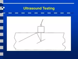
Standard Brachytherapy Procedures Needle Pre-Loaded with Iodine25 Seeds Bladder
Prostate Brachytherapy • Used to treat early localised prostate cancer tumours • Utilises implantation of radioactive seeds • Seeds deliver a local high-dose radiation that decreases • Less damaging than other irradiation techniques as the seeds are implanted directly into the prostate
3-D Ultrasound Method • Sound Waves sent and received at different angles • Returning echoes are processed by a computer which reconstructs a 3-D image • Works much like a CT scan that utilises multiple x-rays (true image)
Image Quality vs. Real-Time • There is a trade-off between quality and how fast the image is processed. • Why is this important?
The Problem • Needles are guided parallel to TRUS transducer • 5mm increments limit lateral positioning • Prostate changes position during procedure • Need constant adjustment, Labour intensive for physician • Enlarge Prostates – sections occluded by patients pubic arch • Parallel insertion completely miss these areas and lead to insufficient dose coverage
Prostate Phantom Testing • Allows for testing of brachytherapy procedures • Prostate mold is cast from a stereo-lithograph of a real patient • Mold injected with molten agar and left to cool (Clear) • Background agar is created to fill the rest of the box (Opaque)
The Solution • Image-guided robotics receiving real-time updates • Removes problem of pubic arch interference • Previous Attempts have distinct advantages • Respective procedures are still invasive • Current project utilises physician • Robot loads, and positions needle Needle Needle
Robot Accuracy • Evaluation of Mean Target Accuracy involves: • Guide the needle to a 3D target • Record the location of the needle within the image
Robot Accuracy • Needle accuracy of actual target vs. “true” measurement and measurement using 3D-guided Robotics
Future Work • Testing on human subjects • Give a better feel for different sized prostates and accuracies • Reduce the size of the robot • Portability, Improve physician accuracy • Improve design to work with larger angles • Increases success of brachytherapy especially in patients with enlarged prostates
Acknowledgements • Aaron Fenster - Supervisor • Adam Krasinski- Supervisor • Jeffrey Bax • David Smith • Laura Bartha • Jacques Montreuil • Shi Sherebrin • Lori Gardi • ChandimaEdirisinghe
References • Bax J, Fenster A, Montreuil J, Gardi L, Smith D. Apparatus And Method For Guiding Insertion of a Medical Tool. US Patent Application No: 11/427,121: Filed June 28, 2007 (Pending). • Fenster A, Downey DB, Cardinal HN. Three-dimensional ultrasound imaging. Physics in Medicine & Biology. 2001 ;46R67-R99. • D.W. Rickey, P.A. Picot, D.A. Christopher, and A. Fenster, “A Wall-less Vessel Phantom for Doppler Ultrasound Studies,” Ultrasound in Med. & Biol 21(9), pp.1163-1176, 1995.