Download
1 / 1
10 likes | 78 Views
Analysis (Fig. 3 - 5 ):. Fig. 3: Fitting of the sections of the fMRI data set into the anatomic 3D data set. Controls. Controls. Patients. Patients. Fig. 8: In patients, beeps and bell ringing additionally stimulate the periaqueductal gray matter.

E N D
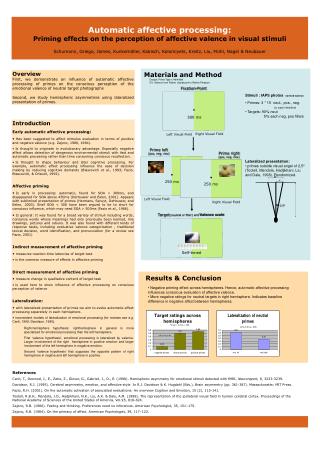
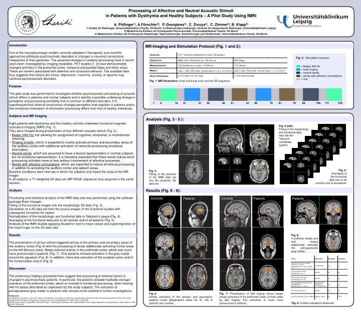
Analysis (Fig. 3 - 5 ): Fig. 3: Fitting of the sections of the fMRI data set into the anatomic 3D data set. Controls Controls Patients Patients Fig. 8: In patients, beeps and bell ringing additionally stimulate the periaqueductal gray matter. Fig. 7: Presentation of bell ringing versus beeps shows activation of the prefrontal cortex on both sides by bell ringing. The activation is much more pronounced in patients. Results (Fig. 6 - 9): Processing of Affective and Neutral Acoustic Stimuli in Patients with Dysthymia and Healthy Subjects – A Pilot Study Using fMRI A. Pöllinger1), A.Förschler2), P. Georgiewa3) , C. Zrouya4), C. Zimmer2), B. Klapp3) 1) Institut für Radiologie, Universitätsklinikum Charité, HU-Berlin; 2) Abteilung Neuroradiologie, Zentrum für diagnostische Radiologie, Universitätsklinikum Leipzig 3) Medizinische Kliniken mit Schwerpunkt Psychosomatik, Universitätsklinikum Charité, HU-Berlin 4) Medizinische Kliniken mit Schwerpunkt Hepatologie, Gastroenterologie, Endokrinologie und Stoffwechsel, Universitätsklinikum Charité, HU-Berlin Introduction One of the neurophysiologic models currently adopted in therapeutic and scientific approaches attributes psychosomatic disorders to changes in neuronal connections, irrespective of their generator. The assumed changes in cerebral processing have in recent years been investigated by imaging modalities. PET studies [1, 2] have demonstrated changed activities in the prefrontal cortex, temporal and parietal lobes and other areas. These are centers associated with attentive and conscious behavior. The available data thus suggests that factors like stress, depression, insomnia, anxiety, or spasms may reinforce psychosomatic disorders. Purpose This pilot study was performed to investigate whether psychosomatic processing of acoustic stimuli differs in patients and normal subjects and to identify a possible underlying change in perceptive and processing sensibility that is common to different disorders. It is hypothesized that external strain/stress changes perception and cognition in patients and/or that emotional modulation of information processing differs from that of healthy individuals. Subjects and MR Imaging Eight patients with dysthymia and five healthy controls underwent functional magnetic resonance imaging (fMRI) (Fig. 1). They were imaged during presentation of four different acoustic stimuli (Fig. 2): Baseline conditions were intervals in which the subjects only heard the noise of the MR imager. In all subjects, a T1-weighted 3D data set (MP RAGE sequence) was acquired in the same session. Analysis Processing and statistical analysis of the fMRI data sets was performed using the software package Brain-Voyager: Fitting of the functional images into the morphologic 3D data (Fig. 3). Generation of a 4D data set from the source images of the functional studies with subsequent correction for motion. Normalization of the morphologic and functional data in Talairach’s space (Fig. 4). Averaging of the functional data sets of all controls and of all patients (Fig. 5). Analysis of the fMRI studies applying Student’s t-test to mean values and superimposition of the result maps on the 3D data sets. Results The presentation of all four stimuli triggered activity of the primary and secondary areas of the auditory cortex (Fig. 6) with the processing of words additionally activating frontal areas on the left (Broca’s area). Beeps induced activity in the prefrontal cortex, which was much more pronounced in patients (Fig. 7). Only patients showed activation in the gray matter around the aqueduct (Fig. 8). In addition, there was activation of the occipital cortex and of the frontomedian area 6 (Fig. 9). Discussion The preliminary findings presented here suggest that processing of external stimuli is changed in psychosomatic patients. In particular, the patients showed markedly stronger activation of the prefrontal cortex, which is involved in emotional processing, when hearing 440-Hz beeps described as unpleasant by the study subjects. The activation of periaqueductal gray matter in patients only remains to be clarified in further investigations. References: [1] Sankaya A, Karasin E, Cermik TF, Abay E, Berkarda S. Evaluation of Dysthymic Disorder with Technetium-99m Hexamethypropylene Amine Oxime Brain Single Photon Emission Tomography. European Journal of Nuclear Medicine 1999; Vol 26; 3:260-264. [2] Thomas P, Vaiva G, Samaille E, Maron M, Alaix C, Steinling M Goudemann M. Cerebral Blood Flow in Major Depression and Dysthymia. J Affect Disord. 1993; Vol 29; 12:235-242 MR-Imaging and Stimulation Protocol (Fig. 1 and 2): Fig. 2: Stimulation protocol. = Beeps 440 Hz = bells ringing = neutral words = words with affective connotations = rest Fig. 1: MR-Parameters of the functional scan and the 3D-sequence. 1 6 16 21 31 36 46 51 61 66 76 81 91 96 106 111 120 Fig. 4 (left): Fitting of the morphologic and functional data sets into the Talairach coordinate system. • Beeps (440 Hz) not allowing for assignment of cognitive, emotional, or motivational meaning. • Ringing of bells, which is expected to mainly activate primary and secondary areas of the auditory cortex with additional activation of networks processing emotional information. • Neutral words, which are assumed to have a lexical representation in normal subjects but no emotional representation. It is therefore expected that these words induce word processing activities more or less without involvement of affective processes. • Words with affective connotations, which are expected to induce emotional processing in addition to activating the auditory cortex and speech areas. Fig. 5: Averaging of the functional data sets of all controls and of all patients Patients Fig. 6: Similar activation of the primary and associative auditory cortex (Brodmann’s areas 22, 41, 42) in patients and controls. Fig. 9: Further activations observed.