Download
1 / 42
420 likes | 430 Views
Reproductive System. Male Reproductive System. Scrotum. A fleshy pouch suspended inferior to the perineum, anterior to the anus and posterior to the base of the penis. Consists of a thin layer of skin and the underlying superficial fascia.

E N D
Scrotum • A fleshy pouch suspended inferior to the perineum, anterior to the anus and posterior to the base of the penis. • Consists of a thin layer of skin and the underlying superficial fascia. • The dermis contains a thin layer of smooth muscle called the dartos, whose resting tone produces the typical wrinkling of the scrotal surface. • A layer of skeletal muscle, the cremaster lies deep to the dermis, its contraction during sexual arousal or in response to cold temperature tenses the scrotum and pulls the testes closer to the body.
Scrotum • The function of the scrotum consists in maintaining the testes at the optimal temperature, around 1.1⁰C (2⁰F) lower than the body’s core temperature to favor the development of spermatozoa
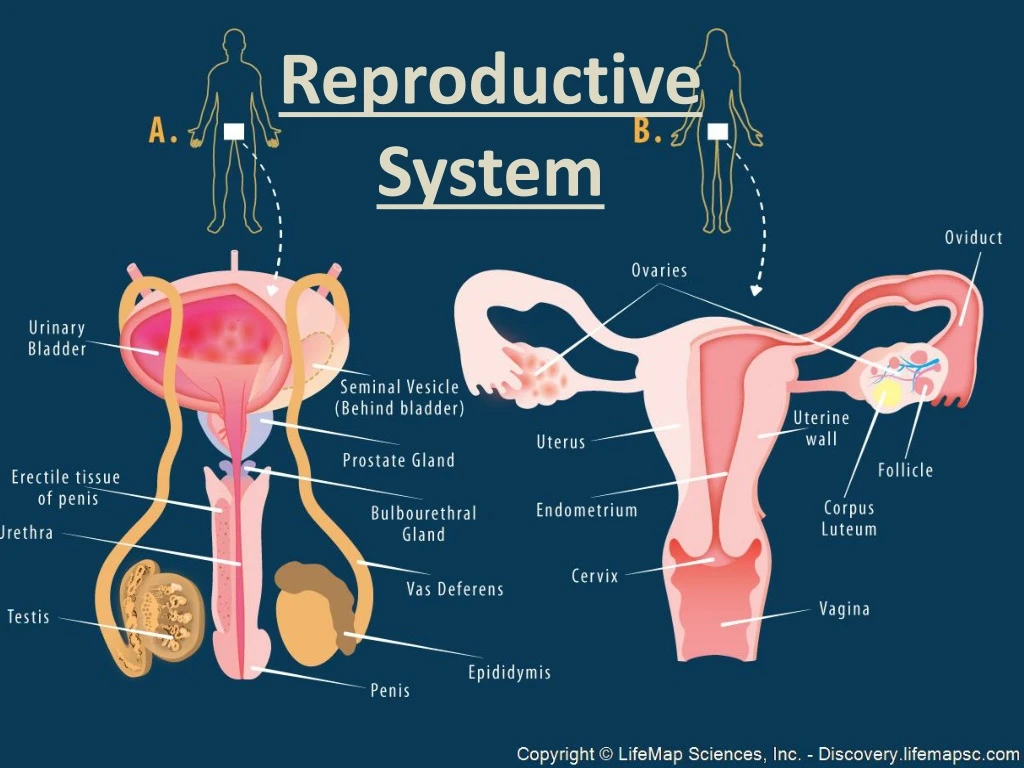
Male External Genitalia Scrotum Testis
Testes • Each testis is about 5 cm long, 3 cm wide and 2.5 cm thick • Each has a weight of around 10-15 g. The left testis is often slightly bigger and lies at a lower position than the right one • The tunica albuginea, a dense layer of connective tissue covers each testis. Its collagen fibers intermesh with those of the epididymis and extend deep into the testis, forming partitions or septa that subdivide the testis into lobules • Distributed among the lobules there are slender, tightly coiled seminiferous tubules. Each tubule is around 80 cm long and each testis contains around one half mile of tubule. Inside these tubules spermatogenesis (sperm cell formation) occurs
Testes • In the spaces between the seminiferous tubules there are small clusters of Interstitial cells (Leydig’s cells) that secrete androgens, the male sexual hormones. • Testosterone is the predominant androgen. • Inside the tubules there are large Nurse cells, also named sustentacular or Sertoli’s cells; productors of the hormonal factor Inhibin a regulator of spermatogenesis. Other products secreted by the nurse cells participate as promoters of sperm cells production an regulators of the embryonic development and differentiation of the male genitalia.
The Male Reproductive Tract • Each seminiferous tubule forms a loop that connects to a network of passageways named the rete testis. • Fifteen to twenty efferent ductulesconnect the distal end of the rete testis to a coiled tubule bound to the upper posterior border of each testis, the epididymis. • The epididymis is around 7m long and is so tightly coiled that it occupies a mere 3 cm behind the testis. • Formed of a head, its part proximal to the testis, a body and a tail, the part that makes continuation with the ductus deferens, the epididymis is an ample parking lot for the storage of spermatozoa
The Ductus (Vas) Deferens • Each ductus deferens or vas deferens is 40-45 cm long, it begins at the tail of the epididymis and as part of the spermatic cord ascends through the inguinal canal entering the abdomen. • It goes curving along the lateral surface of the urinary bladder toward the superior-posterior margin of the prostate gland. • Just before the vas deferens reaches the prostate gland and seminal glands, its lumen enlarges forming the ampulla of the vas deferens • Peristaltic contractions of the vas deferens conduct sperm cells and seminal fluid toward the prostate gland. Sperm cells can be also stored in the vas deferens for several months, during this time the spermatozoa are in a state of “suspended animation” and have a very low metabolic rate
Seminal Glands • Seminal glands are extremely active secretory glands; their secretion contains high amounts of fructose, an energy source for sperm cells; prostaglandins that stimulate smooth muscle contraction along the male and female reproductive tract and fibrinogen that promotes the temporary coagulation of the semen deposited in the female vagina. • Seminal gland secretions are slightly alkaline to neutralize the acid pH of the vagina and their contact with the spermatozoa usually activates their flagellar beating making the male gametes active mobile cells.
Prostate Gland • A small muscular rounded organ about 4 cm in diameter • It encircles the proximal portion of the urethra as it leaves the urinary bladder • The glandular tissue consists of a cluster of 30-50 tubulo-alveolar glands wrapped in a thick layer of smooth muscle fibers. • Its secretion is alkaline and contains mainly seminalplasmin a substance that has an antibiotic effect to help prevent male urinary tract infection • Prostatic secretions are ejected into the prostatic urethra by peristaltic contractions of the smooth muscle in the prostate wall
Bulbo-Urethral Glands • Also called Cowper’s Glands, are situated at the base of the penis, embedded in the fascia of the urogenital diaphragm • They are spheric structures with a diameter of around 10 mm • Their ducts travel alongside the penile urethra for 3-4 cm before emptying into the urethral lumen • They secrete a thick, alkaline mucous secretion that neutralizes urinary acids and also lubricates the glans penis and the urethra; this secretion shows at the urethral opening during the phase of sexual arousal.
Semen • The ejaculate volume is around 2 to 5 mL. • Each mL of semen carries 20 million to 100 million sperm cells • Seminal glands secretion accounts to 60% of the seminal volume • Prostate Gland secretion contribution is around 30% of the seminal volume. • Secretions of the epididymis and the testicular nurse cells are around 5% of the seminal volume • Bulbo-urethral glands secretions are less than 5% of the seminal volume
The Penis • The male copulatory organ is a tubular structure formed of three regions • The crus or root is the fixed portion that attaches the penis to the body wall • The body or shaft is the cylindrical and longer portion of the organ • The glans is the expanded end that surrounds the urethral orifice • A fold of skin called the prepuce or foreskin, covers the glans • Muco-sebaceous glands underneath the prepuce produce a secretion called smegma for lubrication of the glans.
The Penis • Most of the body of the penis consists of three cylindrical columns of erectile tissue: • The two corpora cavernosa run side to side along the dorsal aspect of the penis; they consist of intricate, spaces surrounding a central artery whose fenestrated wall communicates with the spaces and hence can fill them with blood engorging and distending the corpora leading to the penile erection • The single, ventral cylinder, named corpus spongiosumis smaller in diameter and surrounds and protects the penile urethra. It expands at its distal end forming the glans of the penis
Penile Erection • The parasympathetic innervation of the penile arteries involves neurons that release nitric oxide (NO) at their synaptic knobs. • NO relaxes the smooth muscle in the arterial walls and the vessels dilate, blood flow increases and the vascular spaces in the corpora cavernosa engorge with blood leading to penile erection. • The dorsal veins that drain the penis are constricted by the expansion of the corpora cavernosa and this maintains the penis erect. • Orgasm and ejaculation are sympathetic mediated responses. After they occur, it ceases the release of NO and this leads to penile detumescence as blood drains out of the penis.
Urethra • In males, the urethra serves both the urinary and reproductive systems. It extends from the neck of the urinary bladder to the tip of the penis with a longitude of 18-20 cm. It comprises three portions: • The prostatic urethra, goes through the center of the prostate gland • The membranous urethra, the short segment that crosses the urogenital diaphragm, the muscular floor of the pelvis • The spongy urethra or penile urethra that goes along the longitude of the penis, it expands to form the navicular fossa at the glans of the penis then opening at the glans tip at the external urethral orifice.
Female Pelvis (sagittal section) ureter uterine fundus common iliac artery & vein uterine cavity recto-uterine pouch (cul de sac/Douglas’ sac) uterine tube vesico-uterine pouch posterior fornix urinary bladder urethra cervix mons pubis rectum clitoris labium minus vagina labium majus anus introitusvaginalis
Oogenesis • Mitosis of oogonia during fetal life produces a diploid primary oocyte • Meiosis I begins before birth, it won’t be completed until the girl reaches puberty. • Meiosis I is completed after puberty, it produces a secondary oocyte and a first polar body, both are haploid cells. • Secondary oocyte is ovulated in metaphase of meiosis II, process will be completed if fertilization occurs; at completion of meiosis II the mature gamete (a haploid cell) is ready to fuse with the male gamete (also a haploid cell); a second polar body is produced. Fertilization will produce a diploid cell, the Conceptusor Zygote.
Ovulation • Each almond-sized ovary hosts 100.000 to 200.000 primordial follicles • During each ovarian cycle several of these primary follicles develop into secondary follicles • Eventually one secondary follicle develops into a tertiary follicle a.k.a. “Graafianfollicle” “containing an oocyte. • The follicular sac ruptures releasing the oocyte (ovum), this event called “Ovulation” • After ovulation the empty follicle becomes a gland called Corpus Luteum that secretes progesterone and also moderate amounts of estrogen. Progesterone plays an important role in the preparation of the uterus for pregnancy • If the oocyte is not fertilized, the corpus luteum becomes an atretic structure named Corpus Albicans; the internal lining of the uterus (endometrium) sheds and is eliminated as part of the menses.
Oviducts • The two uterine tubes called oviducts or Fallopian tubes have at their lateral ends fingerlike projections called fimbriae • These fimbriae sweep over the surface of the ovary and trap the ovum during ovulation bringing it toward the slightly expanded portion of the oviduct, called infundibulum • Once inside the oviducts, the ovum is moved toward the uterus by the tube peristalsis and the action of the cilia in the tubal lumen. • The oviducts widen along their length in the ampulla and then narrow at the isthmus, at theirpoint of entrance to the uterus • Fertilization of the ovum usually occurs at the distal third of the tubes, the conceptus is then pushed toward the uterus.
Uterus • A pear-shaped muscular sac, located between the urinary bladder and the rectum. • It is around 7.5 cm long and a maximum diameter of 5 cm and weights around 30-40 g • It consists of three major regions : • The fundus, the superior portion, located right over the level of entrance of the oviducts • The body, the most extense portion and the elongated cervix, its portion that shows at the vagina • In its normal position the uterus tilts anteriorly maintaining its cavity mostly aligned with the cervical canal and covering most of the superior and posterior portions of the urinary bladder. This usual bending uterine attitude is called anteversion and it is considered the normal position of the uterus.
Uterine Positions Anteverted (usual, “normal”) Retroverted:Abnormal, prone to prolapse
Female GenitaliaAnterior view A C D A Uterine (Fallopian) tube Broad ligament Uterine fundus Uterine cavity Uterine body Uterine isthmus (segment) Uterine cervix Vaginal lateral fornices Introitusvaginalis External Os of cervix Cervical canal Internal Os of cervix B E B F L K G J Vagina H I
Uterus • The tubular cervix projects into the vagina, the vaginal folds around the cervix are called each one a vaginal fornix. • The uterine body is the broadest part of the uterine cavity, the usual site for the implantation of the placenta during pregnancy and the area where the developing fetus resides.
Uterus: Suspensory Ligaments • A double layered fold of peritoneum called the mesovarium holds the ovaries to the broad ligament of the uterus • The suspensory ligaments hold the ovaries to the pelvic wall • The ovarian ligaments hold the ovaries to the uterus • The round ligaments extend laterally from the ovaries and provide posterior support.
The Uterine Wall • The uterine wall consists of three main tissue layers: • The perimetrium, the outer layer, is an extension of the visceral peritoneum and hence called a serosa. • The myometrium is the thick middle layer, composed of three layers of smooth muscle, responsible for the powerful contractions during labor • The endometrium is a double epithelial layer, its inner basilar zone produces a new functional zone each month. The outer functional zone is a very active glandular tissue able to support an implanted embryo. It sheds on every cycle and is discarded as part of the menses
Vagina • An elastic, distensible muscular tube, around 7.5 -9 cm long extending from the cervix to the vestibule, a space bounded by the female external genitalia. • The shallow recess surrounding the protruding cervix is called the vaginal fornix • The vagina lies parallel to the rectum which is posterior to her. Anteriorly, the urethra extends along the superior end of the vagina to open in the area of the vestibule • The vagina has three major functions: • Serves as a passageway for the menses • Is the female copulatory organ • Forms the inferior portion of the birth canal
Female External Genitalia • The vulva includes the following structures: • Mons Pubis, a pad of adipose tissue over the pubic symphysis, usually covered by pubic hair • The labia majora, (singular: labium majus), two longitudinal folds of skin, extending from the pubis to the posterior angle of the vulva • The labia minora, (singular: labium minus), two small folds of skin covered internally by a mucosa, they extend longitudinally and guard the vestibule, the place where the vagina opens. • The clitoris, is a small cylindrical mass of erectile tissue, the analogous of the penis; it also has a prepuce covering its distal tip called the glans • The entrance to the vagina, the introitusvaginalisis often covered by an elastic epithelial fold called the hymen
Female Mammary Gland • The female mammary glands are modified sweat glands. They complete their anatomical development at puberty, under the stimulus of estrogens. • Fat deposition is the main contributor to the size of the breast, and its size is not related with the capacity of the organ to produce milk for lactation of the offspring. • Each gland consists of 15 to 20 lobes of glandular tissue separated by fat and connective tissue. Each lobe contains smaller lobules formed of milk-secreting cells called alveoli. • Lactiferous ducts drain milk from the lobules toward the lactiferous sinuses. These sinuses empty the milk to a raised portion of the breast called the nipple. • A circular pigmented area of skin called the areola surrounds the nipple.