Download
1 / 36
360 likes | 385 Views
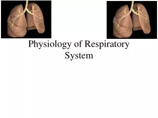
Physiology of respiratory system. External breathing. General functions of respiratory system.

E N D
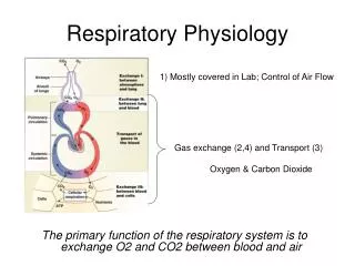
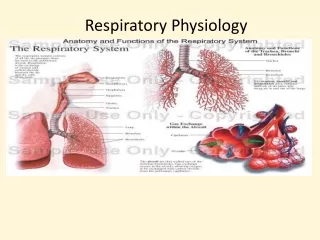
General functions of respiratory system • The respiratory system comprises of the nose, mouth, throat, larynx, trachea, bronchi and lungs. The function of the respiratory system is to facilitate gaseous exchange to take place in the lungs and tissue cells of the body. • Oxygen is required by cells in the body to allow various metabolic reactions to take place and to produce energy and is therefore essential to life. The respiratory system may be defined as the organs and tissues through which air is passed into and out of the body to allow the necessary gaseous exchanges to take place.
Respiratory System - a complex of structures that participate in gas exchange, and mechanisms for their regulation.The activity of respiratory system is best evaluated by oxygen consumption (OC) in 1 min. In the adult resting OC is about 3.5 ml (min / kg).
“Respiration” includes three separate but related functions: • (1) ventilation (external breathing); • (2) gas exchange, which occurs between the air and blood in the lungs and between the blood and other tissues of the body; • (3) oxygen utilization by the tissues in the energy – internal breathing
Stages of exchange of gases }ventilation • 1. exchange of gases between atmospheric air and intraalveolar air • 2. exchange of gases between intraalveolar air and blood • 3. transport of gases • 4. exchange of gases between blood and tissue • 5. internal (tissue) respiration }diffusion }perfusion
Respiratory muscles • Main muscles for inspiration: diaphragm and external intercostal, intercartilaginous muscles • Muscles for expiration: internal intercostal, pectoral, abdominal muscles
Muscles of Inspiration: Diaphragm Most Important Muscle Of Inspiration • Responsible for 75% of inspiratory effort • Thin dome-shaped muscle attached to the lower ribs, xiphoid process, lumbar vertebra • Innervated by Phrenic nerve (Cervical segments 3,4,5) • During contraction of diaphragm • Abdominal contents forced downward & forward causing increase in vertical dimension of chest cavity • Rib margins are lifted & moved outward causing increase in the transverse diameter of thorax • Diaphragm moves down 1cm during normal inspiration • During forced inspiration diaphragm can move down 10cm • Paradoxical movement of diaphragm when paralyzed • Upward movement with inspiratory drop of intrathoracic pressure • Occurs when the diaphragm muscle is denervated
Inspiration • The dome of the diaphragm flattens, ribs elevate • In the rest, 4/5 of inhalational work is done by diaphragm. • Pressure in the alveoli reduces below atmospheric, the air moves under pressure gradient into the lungs
Biomechanism of breathing • Breathing is an active process - requiring the contraction of skeletal muscles. The primary muscles of respiration include the external intercostal muscles (located between the ribs) and the diaphragm (a sheet of muscle located between the thoracic & abdominal cavities). • The external intercostals plus the diaphragm contract to bring about inspiration: • Contraction of external intercostal muscles > elevation of ribs & sternum > increased front- to-back dimension of thoracic cavity > lowers air pressure in lungs > air moves into lungs • Contraction of diaphragm > diaphragm moves downward > increases vertical dimension of thoracic cavity > lowers air pressure in lungs > air moves into lungs: To exhale: • relaxation of external intercostal muscles & diaphragm > return of diaphragm, ribs, & sternum to resting position > restores thoracic cavity to preinspiratory volume > increases pressure in lungs > air is exhaled
Exhalation • Normally is a passiveprocess. After relaxation of muscles,due to the elastic tension of thorasic tissues air is removed (Becoming active duringbronchial obstruction)
The Pleura Space • Two parts of the pleural membrane • Visceral pleura is a thin serosal membrane that envelopes the lobes of the lungs • Parietal pleura lines the inner surface of the chest wall, lateral mediastinum, and most of the diaphragm • Pleura space enclosed by a continuous membrane • The two pleural membranes slide against each other • The pleural membranes are difficult to separate apart • Separated by a thin layer of serous fluid ( a large amount would be a pleural effusion as seen in CHF, CA, infection) • Pleura sac • The continuous membranes fold to create a sac inferiorly • Both pleura line this potential space inclosing a small amount of fluid • Pleural fluid • Functions as a lubricant between the membranes, prevents frictional irritation • Causes the visceral & parietal pleura to adhere together, maintains surface tension • Lymphatic drainage maintains constant suction on pleura (-5cmH2O)
Pleural Pressure • The pressure of the fluid in the space between the lung pleura (visceria) & chest wall pleura (parietal), always negative • Normally at rest suction creates a negative pressure at beginning of inspiration (-5cmH20) • This suction holds the lungs open at rest • Pressure becomes more negative during inspiration moving to -7.5cmH20 allowing for negative pressure respiration • If pleural pressure becomes positive the lung will collapse: Pneumothorax, Hemothorax, Chylothorax
Pressure in the lungs and intrapleural pressure • Intrapleural pressure is always lower than the alveolar one:First: chest is a sealed container.Second, the lungs are characterized by elastic tension, which is due to these factors: • 1. presence of ellastic fibers, which make 1 / 3 of elastic tention;2. surface tension of the liquid layer on the inner surface of alveoli, which makes 2 / 3 of the elastic tension of the lungs. • Thirdly, “negative” pressure in the pleural cavityis maintained by the large absorbtion capacity of pleural leaves.
Pressure in lungs • As the external intercostals & diaphragm contract, the lungs expand. The expansion of the lungs causes the pressure in the lungs (and alveoli) to become slightly negative relative to atmospheric pressure. • As a result, air moves from an area of higher pressure (the air) to an area of lower pressure (our lungs & alveoli). • During expiration, the respiration muscles relax & lung volume descreases. This causes pressure in the lungs (and alveoli) to become slight positive relative to atmospheric pressure. As a result, air leaves the lungs.
Lung capacity • 1. The total maximum lung capacity - the maximum volume of air that fits in the lungs or the sum of all lung volumes. Normally is 4,5-6,5 liters. • 2.Vitallung capacity - the largest amount of air you can exhale after maximum inhalation or the amount of the first three volumes. Normally it is: women - 3,0-3,5 l; in men - 3,5-5,0 liters. • 3. Inspiratory capacity - the maximum amount of air you can breathe after calm exhalation, or the amount of the first two volumes. Normally it - 1,8-2,8 liters. • 4. Functional residual capacity - the amount of air contained in the lungs after calm exhalationor the amount of the last two volumes. Normally - 2.5-3.5 liters.
REMEMBER: Spirometry cannot measure Residual Volume (RV) thus Functional Residual Capacity (FRC) and Total Lung Capacity (TLC) cannot be determined using spirometry alone. • FRC and TLC can be determined by 1) Helium dilution, 2) Nitrogen washout, or 3) body plethysmography