Download
1 / 55
550 likes | 555 Views
THE N.S. The nervous and endocrine systems control the functions of the body. FUNCTIONS. (1) It receives information ( stimuli ) that arise either outside or inside the body from different sensory receptors. These are grouped together to form sensory input. FUNCTION.

E N D
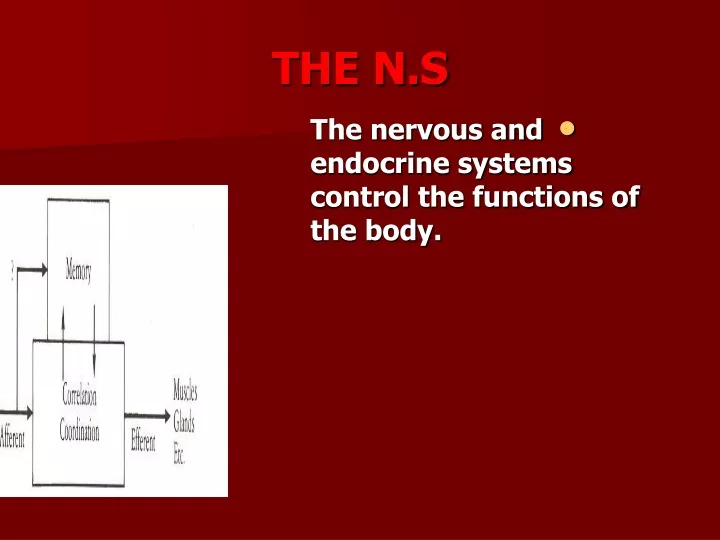
THE N.S • The nervous and endocrine systems control the functions of the body.
FUNCTIONS • (1) It receives information (stimuli) that arise either outside or inside the body from different sensory receptors. • These are grouped together to form sensory input.
FUNCTION • (2) It integrates andcorrelatesthese sensoryinput. • (3) It effects a response to an effector (muscle or gland) through the motor output.
CLASSIFICATION OF N.S. • (1) Central nervous system(CNS) • (2) Peripheral nervous system (PNS)
CENTRAL NERVOUS SYSTEM • It is formed of : • The brainand spinal cord. • They occupy the dorsal body cavity. • They are the main centers where correlation and integration of nervous information occur.
PERIPHERAL N.S • It is the link between the CNS and peripheral structures. • It is formed of : • 1.Cranial and Spinal nerves. • 2.Their ramifications within the body. • 3.Associated Ganglia.
CLASSIFICATION OF NERVES • Nerves are classified according to the direction in which they carry impulses : • (1) sensory (afferent): • They carry impulses toward the CNS. • (2) Motor (efferent) : • They carry impulses from the CNS. • (3) Mixed .
FUNCTIONAL CLASSIFICATION OF PNS • 1.Sensory (Afferent): • A.Somatic : • It delivers impulses to the CNS from: • Skin. • Skeletal muscles. • joints. • B. Visceral : • It transmits impulses from the visceral organs.
FUNCTIONAL CLASSIFICATION OF PNS • 2. Motor (efferent ) • A. Somatic : • It allows conscious (voluntary) control of skeletal muscles. • B. Autonomic • It controls involuntary activity of smooth, cardiac muscles and glands.
AUTONOMIC N.S • It is divided into Sympathetic and Parasympathetic. • In both there are afferent, efferent nerve fibers and ganglia.
AUTONOMIC N. SYSTEM • It has a chain of two motor neurons (Ganglia). • The first motor neuron is in the brain or spinal cord. • Its axon (Preganglionic)synapse with the second motor neuron (outside ) the CNS. • The axon of this neuron is (Postganglionic) extends to the organ which it serves.
INTERNAL STRUCTURE OF N.S. • The interior of the N.S. is organized into gray and white matter.
GREY MATTER • It is composed of large numbers of excitable nerve cells (Neurons) and their processeswhich are supported by specialized tissue (Neuroglia).
NEURONE • It is the structural and functional unit of the nervous system. • It receives and integrates information from sensory receptors or other neurons and transmit information to the effector organs.
PROCESSES OF NEURONS • Dendrites : • They are receptive processes. • They can detect changes in the external or internal environment.
PROCESSES OF NEURONS • Axons : • Carry information away from the cell body. • They can be divided into several collaterals. • Terminal Buttons : • Are at the end of the axon where information is transferred to the dendrites of other neurons. Transmission of information between neurons is always by chemical not electrical means.
FUNCTIONAL CLASSIFICATION OF NEURONS • 1. Sensory • Carry information from the periphery to the CNS to reach the conscious level. • Their cell bodies are always outside the CNS in a ganglion.
2. MOTOR (EFFERENT) Their cell bodies are always located in the CNS. • They are divided into upper and lower motor neurons.
3. ASSOCIATION (INTERNEURONS) • They are relay neurons • connecting sensory and motor neurons. • They reside entirely within the CNS. • Their cell bodies are always located in the CNS.
WHITE MATTER • It consists of nerve fibers embedded in neuroglia. • Tracts : • Nerve processes sharing common connections, course and functions.
REFLEXES • They are rapid, predictable and involuntary responses to stimuli. • They are : • (1) Somatic reflexes : • Stimulate skeletal muscles. • (2) Autonomic reflexes : • Regulate smoothmuscles.
PROTECTION OF CNS • The brain and spinal cord are covered with meninges and are suspended in the cerebrospinal fluid. • They are further protected by the bones of the skull and vertebral column.
BRAIN • Is divided into : • Fore brain. • Mid brain. • Hind brain.
HIND BRAIN • It consists of : • Cerebellum • Medulla . • Pons.
MEDULLA OBLONGATA • It is continuous caudally with the spinal cord. • It extends rostrally to the pons.
PONS • It owes its name to its appearance as that of a bridge connecting the two cerebellar hemispheres.
CEREBELLUM • It is an important center which is essential for accurate, coordinated and purposeful movements. • It operates at an unconscious level.
STRUCTURE • It has an outer layer of grey matter (cerebellar cortex) which is highly convoluted and forms regular folds (Folia). • The white matter forms the central core.
MID BRAIN • It is rostral to the pons. • Its dorsal surface is formed by four rounded eminences(Superiorand InferiorColliculi).
BRAIN STEM • It forms a small part of the entire brain. • It consists of : • Medulla. • Pons. • Mid brain (It is the smallest part of the brain stem).
BRAIN STEM • It is attached to the cerebellum by three parts of nerve fibers (peduncles) : • Inferior to the medulla. • Middle ( largest peduncle) to the pons. • Superior to the mid brain.
FORE BRAIN It consists of : Diencephalon . Two cerebral hemispheres.
DIENCEPHALON • It is formed of four subdivisions in a dorsoventral direction • (1) Epithalamus • The Pineal Gland is its most notable part . • It is immediately rostral to the superior colliculi of the mid brain.
DIENCEPHALON • (2) Thalamus • It is the largest part. • It has an important role in the sensory, motor and cognitive functions.
DIENCEPHALON • (3) Subthalamus • .(4) Hypothalamus • It is involved with the autonomic, limbic and cardiovascular systems.
CEREBRAL HEMISPHERES • They represent the massive part of the forebrain. • The two hemispheres are separated by theGreat Longtudinal Fissure. • The fissure is occupied by theFalx Cerebri.
CEREBRAL HEMISPHERES • The cerebral cortex is highly convoluted for maximising the cortical surface area. • The convolutions are the Gyri and the furrows between them are the Sulci. • Some of these gyri and sulci mark the location of important functional areas.
CEREBRAL LOBES • Each hemisphere is divided into four lobes which are named according to the names of the bones of the skull beneath which they lie.
CEREBRAL LOBES • The lobes are : • Frontal lobe . • Its most anterior convexity is the frontal pole. • Parietal lobe. • Temporal lobe. • Its tip is the temporal pole. • Occipital lobe. • It terminates in the occipital pole.
STRUCTURE • It consists of an outer layer of grey matter (cortex) and an inner mass of white matter.
BASAL GANGLIA • They are large masses of cell bodies (grey matter) which are buried within the white matter.
BASAL GANGLIA • They are concerned with the control of muscle tone posture.They are important in the facilitation of appropriate motor behavior and the inhibition of unwanted movements.
BASAL GANGLIA • The largest component is the Corpus Striatum. • It consists of : • Caudate nucleus. • Putamenand Globus pallidus.
FUNCTIONAL CORTICAL AREAS(Frontal lobe) • Precentral gyruswhich contains theprimary motor cortex. • It is the highest level in the brain for the control of voluntary, skilled movements(muscles of speech, fascial expression and hand movements). • The opposite half of the body is represented in a highly precise fashion.
UPPER MOTOR NEURONES • They are in the motor cerebral cortex. • Their axons form the descending tracts (corticospinal and corticobulbar) which control the activity of the lower motor neurons.
LOWER MOTOR NEUROENS • Their cell bodies are in the spinal cord and brain stem. • They constitute the final pathway by which the nervous system controls the skeletal muscles.
FUNCTIONAL CORTICAL AREAS(PARIETAL LOBE) • Postcentral gyrus (primary somatosensorycortex) • It is the site of termination of pathways carrying touch, pressure, pain and temperature from the opposite half of the body.
FUNCTIONAL CORTICAL AREAS • Occipital Lobe : • Visual cortex • Temporal Lobe : • Auditory cortex
CORTICAL FIBERS • Corona Radiata • It is a radiating pattern which • is formed from the afferent and efferent fibers between the cerebral cortex and subcortical structures.
CORTICAL FIBERS • Internal Capsule • It is deep inside the hemisphere. The fibers are arranged as a dense sheet of white matter