Download
1 / 25
250 likes | 380 Views
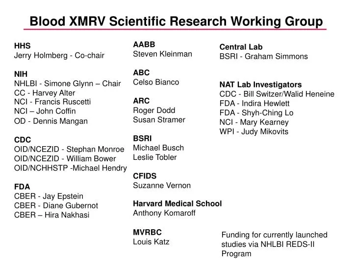
Blood XMRV Scientific Research Working Group. HHS Jerry Holmberg - Co-chair NIH NHLBI - Simone Glynn – Chair CC - Harvey Alter NCI - Francis Ruscetti NCI – John Coffin OD - Dennis Mangan CDC OID/NCEZID - Stephan Monroe OID/NCEZID - William Bower OID/NCHHSTP -Michael Hendry FDA

E N D
Blood XMRV Scientific Research Working Group HHS Jerry Holmberg - Co-chair NIH NHLBI - Simone Glynn – Chair CC - Harvey Alter NCI - Francis Ruscetti NCI – John Coffin OD - Dennis Mangan CDC OID/NCEZID - Stephan Monroe OID/NCEZID - William Bower OID/NCHHSTP -Michael Hendry FDA CBER - Jay Epstein CBER - Diane Gubernot CBER – Hira Nakhasi Central Lab BSRI - Graham Simmons NAT Lab Investigators CDC - Bill Switzer/Walid Heneine FDA - Indira Hewlett FDA - Shyh-Ching Lo NCI - Mary Kearney WPI - Judy Mikovits AABB Steven Kleinman ABC Celso Bianco ARC Roger Dodd Susan Stramer BSRI Michael Busch Leslie Tobler CFIDS Suzanne Vernon Harvard Medical School Anthony Komaroff MVRBC Louis Katz • Funding for currently launched • studies via NHLBI REDS-II Program
Blood XMRV Scientific Research Working Group Mission - design and co-ordinate research studies to evaluate whether XMRV poses a threat to blood safety Working Group includes representatives from transfusion medicine, retrovirology, and CFS scientific communities, as well as representatives from key Federal Agencies including HHS, FDA, CDC and NIH Evaluation of blood safety risks includes several steps:Evaluate XMRV nucleic acid and antibody assays Establish prevalence of XMRV in blood donors Determine if XMRV is transfusion-transmitted Determine if transfusions are associated with CFS or prostate cancer (epidemiology studies)
XMRV SRWG - Study Phases • Phase I - Analytical Panels • Evaluate performance of XMRV NAT assays • Phase II - Pilot Clinical Studies • Whole Blood versus PBMC • Timing of sample preparation • Phase III - Clinical Sensitivity/Specificity Panel • Assay performance on pedigreed clinical samples • Phase IV - Blood Donor Clinical Panel • Initial estimation of XMRV nucleic acid prevalence in blood donors • Initiation of donor seroprevalence studies
Phase I - Analytical Panels • Analytical performance panels • Comparison of Limit Of Detections and accuracy of Viral Loads of current assays; standardize performance of future XMRV detection assays for blood cells and plasma • Whole blood panel - spiked with XMRV positive cells • Plasma panel - spiked with supernatant containing XMRV • 22Rv1 cells • Human prostate cell line chronically infected with XMRV • Contain at least 10 XMRV proviral copies • Knouf et al (2010) J Virol 83:7353 (PMID:19403664) • Virus supernatant • Supernatant from cultured 22Rv1 cells • Approximate Viral Load of 5 x 10⁹ RNA copies/ml
Phase II - Pilot Studies • Facilitate labs with divergent results to test clinically validated samples • Whole Blood versus PBMC versus plasma • Original Lombardi et al study performed on isolated PBMC and plasma • Large scale studies for prevalence facilitated by using WB or plasma • Donor-recipient and other repositories are mainly comprised of frozen WB and/or plasma • Timing of Processing • Processing and freezing of samples for study varied from 2-4 days due to requirements for completion of infectious disease (ID) testing. Other frozen WB and plasma repositories were prepared 1-3 days post-phlebotomy • Studies with other cell-associated viruses (HERVs, HTLV, herpesviruses and anelloviruses), demonstrate that levels of viral nucleic acid in plasma and whole blood vary with time from collection to processing and frozen storage
Phase III - Clinical Sensitivity and Specificity • The ability of participant assays to effectively detect clinical XMRV positive and negatives will be examined • To attempt to overcome the issues of clinical variation and specificity, larger numbers of pedigreed clinical XMRV-positive and -negative samples are being assembled into a coded panel to be distributed to all labs • Positives • 30 clinical samples will be collected by WPI and Harvard. All will be from patients reported by multiple assays as XMRV/MRV+ in the Lombardi et al or Lo et al studies. Method and timing of processing will be determined based on pilot study data • Negatives • PCR detection assays are prone to detect the amplification of non-specific human derived genomic sequences • Thus, a larger group of 10 pedigreed negatives are required in order to introduce generic variability • Will be pedigreed negative by PCR at WPI and CDC labs, and for serology at WPI, CDC and NCI labs (Ruscetti/Bagni)
Phase IV - Clinical Panel for Donor Prevalence • Blinded panels consisting of approximately 300 blood donor samples, as well as confirmed XMRV/MRV-positive and negative analytical standards and clinical samples (from phase III) will be created for WB and plasma • Plan would be to distribute blinded panels to at least 4 of the participating laboratories for nucleic acid & serological testing • Test results will be analyzed: • Correlation between whole blood and plasma testing will be determined for each lab and between labs Preliminary XMRV prevalence in blood donors (proportion of donors positive for XMRV nucleic acids and/or antibodies) will be estimated based on compiled results from each lab
Phase I - Analytical Panel Production Whole blood Plasma Whole Blood (WB) unit from pedigreed (NAT and serology) negative donor Plasma components from two pedigreed (NAT and serology) negative donors 22Rv1 cells spiked into WB to yield 9,900 22Rv1 cells per ml of WB 22Rv1 supernatant spiked to give approximately 250,000 RNA copies per ml of plasma Three-fold dilutions in fresh WB Five-fold dilutions in fresh plasma 0.5 ml aliquots to give 15 panels with three replicates at each dilution, plus 6 negative controls 0.5 ml aliquots to give 15 panels with three replicates at each dilution, plus 6 negative controls
Conclusions and Limitations • XMRV NAT detection assays were highly sensitive • Five assays (CDC, FDA-Lo, NCI, WPI, Gen-Probe) for whole blood and all six assays for plasma were found to have no substantial differences in terms of sensitivity • The overall similarity of results suggests sensitivity of assays cannot explain differences in XMRV detection in clinical samples reported by the participating laboratories • Caveats • XMRV isolate with which 22Rv1 cells are infected may not adequately represent the diversity of XMRV clinical isolates • Use of two units of plasma as diluent for preparation of the spiked plasma panel may have compromised the accurate dilution of virus
Phase IIa - Setup • XMRV-positive samples • WPI collected blood from four subjects (mostly with CFS) previously identified as XMRV positive in the Lombardi et al study (by PCR, serology and/or culture) • Specimens separated into tubes and were processed immediately, or left at 4oC for 2 or 4 days • Each specimen was processed into replicate PBMC, WB and plasma samples that were frozen for panel preparation • Analysis • Unblinded panels were distributed to WPI and CDC and a blinded panel to NCI for testing
Phase II Subject Characteristics • Four Females • 26-53 Years of age • Three were diagnosed with CFS over 20 years ago, one is a family member of a long-time CFS patient
Phase IIa - CDC Results Plasma was ultracentrifuge pelleted prior to nucleic acid extraction All PCR positive samples tested negative for mouse mitochondrial DNA
Phase IIa - WPI Plasma Results Plasma processed using Qiagen Viral RNA kit Whole blood samples were all negative for DNA PBMC were not tested
Phase IIa – NCI/DRP results • Single-Copy quantitative PCR assay for XMRV gag • Plasma spiked with internal control virus and ultracentrifuged prior to nucleic acid extraction using guanidinium isothiocyanate and PCR was performed +/- RT step • WB and PBMC extracted for DNA • All samples (Plasma, WB and PBMC) and all time-points (Day 0, 2, 4) were negative • Plasma internal controls for pelleting of virus and RT step were all in range • WB and PBMC genomic DNA controls in range
Phase IIa - Conclusions and Limitations • Two out of three labs detected XMRV in clinical samples • Plasma out-performed WB in both labs, and PBMC in the one lab that tested PBMC • Day 2 and day 4 samples out-performed day 0 plasma samples in both labs • Caveats • Panel was distributed mostly unblinded • Small sample size • Third laboratory failed to detect virus despite sensitive assay
Phase IIb - Setup • XMRV-positive samples • Blood collected into EDTA tubes by an independent phlebotomist at patients home or work from the same four subject (one patient had independently initiated anti-retroviral therapy) • The pedigreed negative subject used for the analytical panel and phase IIa panel was bled by the same phlebotomist as a control • Phlebotomist directly supplied the samples to BSRI for processing • Samples separated into tubes and were processed the same day, or left at 4oC for 2 days • Each sample was processed into PBMC, WB and plasma • Analysis • Blinded panels were distributed by BSRI to WPI, CDC, NCI and Gen-Probe for testing. Two sets of the panel were retained by BSRI to distribute for follow-up work as needed • Results were reported back to BSRI and decoded
Phase IIb – NCI/DRP results • Single-Copy quantitative PCR assay for XMRV gag • Plasma spiked with internal control virus and either extracted directly or ultracentrifuged prior to nucleic acid extraction using guanidiniumisothiocyanate and used +/- RT step • WB and PBMC extracted for DNA and RNA and used +/- RT step • All Plasma and PBMCs samples at both time points (Day 0, 2) were negative • Plasma internal controls for pelleting of virus and RT step were all in range
Phase IIb – CDC results • Multiple Assays for XMRV and generic MLV • Plasma ultracentrifuged prior to nucleic acid extraction • Plasma assayed with nested RT-PCR for XMRV gag and envelope and quantitative RT-PCR for MLV gag and integrase • WB and PBMC assayed with nested PCR for XMRV polymerase and quantitative PCR for MLV protease • All Plasma and PBMCs samples at both time points (Day 0, 2) were negative • Plasma RNA controls in range • WB and PBMC genomic DNA controls in range
Phase IIb – Gen-Probe results • Target Capture/Transcription-Mediated Amplification (TMA)/chemiluminescent detection • Platform: TIGRIS System • High throughput, fully automated NAT System • Currently used routinely world-wide for NAT blood screening • Assay design: duplex assay that targets conserved sequences in two separate regions of XMRV genome • Ability to detect MRV-like sequences is not known; new assay to detect a broad range of MRV sequences is under development • Each reaction includes an internal control which validates assay steps (target capture, amplification and detection) • Sample volumes: 500 µl plasma, 50 μl WB or 100 μl PBMC • All Plasma and PBMCs samples at both time points (Day 0, 2) were negative
Phase IIb – WPI results • Nested RT-PCR for MRV gag followed by sequencing of positive bands to confirm specificity • RNA extracted directly from plasma • Total nucleic acid extracted from PBMC and RT step performed • Whole Blood has not been completed Negative – Negative by nested PCR/sequencing + - Band of correct band size, sequences as XMRV/MRV a. - Investigation following decoding of results determined that there was a procedural error during PBMC sample extraction involving reuse of needles (employed to lyse cells and shear DNA) on sequential PBMC cell pellets.
Summary of Phase IIb Serology Results NCI – Flow cytometry on cells expressing spleen focus-forming virus env (Lombardi et al, 2009) CDC – Western blot for multiple MLVs (Switzer el, 2010)
In this round of testing, only one out of four labs detected XMRV in clinical samples and only on PBMC Ultracentrifugation or direct extraction of plasma did not seem to make a difference in detection by NCI lab Sensitive NAT assay from a diagnostic company was unable to detect XMRV/MRV Conclusions from Phase IIb
Conclusions • Based on Phase II findings, no clear advantage to delayed processing • Include serology in parallel with NAT in future studies • Continue with collection of Phase III panel • Include more positive and negative samples and differently sourced samples • PBMC, WB, and plasma will be used, with a standard next day processing protocol